Evaluating the First Software Feature for 3D Visualization of Cryoballoon Ablation Catheters in Atrial Fibrillation
Introduction: Pulmonary Vein isolation (PVI) is the standard treatment approach in interventional therapy of atrial fibrillation. There has not been any software support for navigation during cryoballoon ablation procedures, whereas various electroanatomical mapping systems are already available to support 3D navigation for RFCA. We present an entirely X-ray image-based software tool that facilitates 3D visualization and navigation for cryoballoon ablations.
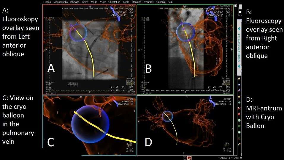
Methods: The new software was evaluated in 90 cryoballoon ablation procedures. A 3D model of the left atrium (LA) was segmented out of a pre-procedural MRI angiography of the left atrium (LA). The LA model is overlayed onto fluoroscopic images and registered to the patient’s anatomy using the new software. Based on a biplane fluoroscopic image, acquired during each cryoballoon freeze, a cryoballoon model was semi-automatically generated, automatically imported into the software and displayed relative to the LA model (Fig. 1).
Results: The cryo balloon reconstruction was successful in 100% of applications (90 ablation procedures with 575 freezes). The mean time for 3D reconstruction and visualization was 3.35 sec ± 0.7 sec.
Conclusions: We present the first software supporting 3D navigation during cryoballoon procedures. It offers intuitive and fast 3D visualization of cryoballoon catheters, a feature that has only been available for RFCA and electroanatomic mapping systems. The new software does not require additional equipment or new hardware. Instead, it uses and combines the existing devices and data in the EP lab using X-ray images only.
Powered by Eventact EMS