
Limited Accuracy of Myocardial Deformation Imaging in Diagnosing Left Ventricular Segmental Dysfunction: is it Only a Limitation of the 2D Strain Software?
2Faculty of Medicine, Technion, Haifa
Background: Two-dimensional strain (2DS) may aid in diagnosing left ventricular (LV) segmental dysfunction, but its accuracy is limited.
Methods: Echocardiographic 2DS imaging was performed in 54 patients (pts) with a first ST-elevation myocardial infarction (MI) and single-vessel disease [age 56±11 years; 74% men; infarct-related coronary artery – left anterior descending artery in 34 pts (63%); LV ejection fraction: 47±10%; wall motion score index: 1.67±0.26]. Peak longitudinal systolic strain (pSS) and systolic strain rate (SR) were measured in 3 apical views (EchoPAC, GE). The echocardiographic “gold standard” for LV segmental function was determined visually by 2 independent experienced cardiologists. Disagreements between these observers were resolved by a third blinded observer.
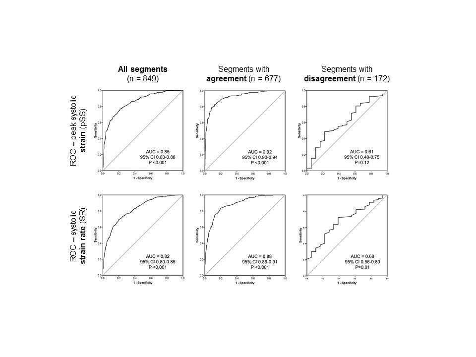
Results: 2DS measurements were feasible in 849 of 864 LV segments (98.3%). By the multiple observers algorithm, 490 (57.7%) segments were classified as normal, 137 (16.1%) hypokinetic, and 222 (26.1%) akinetic. Using receiver operating characteristics (ROC) analysis, the area under the curve (AUC) for detecting segmental abnormality (hypokinetic or akinetic segments) by pSS was 0.85 (95% confidence interval 0.83-0.88) and for detecting akinetic segments was 0.88 (0.85-0.90); the corresponding values for SR were 0.82 (0.80-0.85) and 0.83 (0.80-0.86). The 2 initial observers agreed in classification of 677 (79.7%) segments. When the segments with and without agreement between the 2 initial observers were analyzed separately, the corresponding AUC values were slightly higher for segments with agreement [0.92 (0.90-0.94) and 0.92 (0.90-0.94) for pSS and 0.88 (0.86-0.91) and 0.88 (0.85-0.91) for SR] and much lower for segments with disagreement between the 2 initial observers [0.61 (0.48-0.75) and 0.64 (0.54-0.73) for SS and 0.68 (0.56-0.80) and 0.59 (0.48-0.69) for SR].
Conclusion: Longitudinal pSS and SR can assist in the diagnosis of segmental LV dysfunction. Their limited diagnostic accuracy appears to be partially due to the inherent limitation of visual classification of segmental function.
Powered by Eventact EMS