During recent years it has been demonstrated that carbon nanotubes (CNTs) spontaneously dissolve in chlorosulfonic acid (Davis et al., 2009), and at high concentrations form a liquid crystalline phase (Davis et al., 2004). Actually, chlorosulfonic acid (CSA) is the only solvent for CNT, to form thermodynamically stable solutions and liquid crystalline phases, from which carbon fibers can be spun. Fiber spinning from a liquid crystal state is essential for the high degree of CNT orientation in the fibers, and hence preserving the intrinsic unique properties of individual CNT in the spun fiber (Behabtu et al., 2013).
The transition between the isotropic and the liquid crystalline phases depends strongly on the CNT type, concentration, and solvent strength. Combination of direct cryogenic transmission- and cryogenic scanning-electron microscopy (cryo-TEM and cryo-SEM) of CNT/CSA solutions at different concentrations allowed us, for the first time to follow phase transformation at nanometric level; from diluted solution to the isotropic phase, through the biphasic region, to the pure liquid crystalline phase, used as the "dope" for fiber spinning (See Fig.1). To allow direct imaging of superacid solutions we developed novel cryo-EM specimen preparation and imaging methodologies, suitable for highly acidic systems. Those techniques preserve the native nanostructure in the system, without harming the expensive equipment and the operator (Kleinerman et al., 2015), and were successfully applied to study CNTs (Davis et al., 2009), graphene (Behabtu et al., 2010), and boron nitride nanotubes (Kleinerman et al., in preparation) in CSA in their native state.
The correlation between direct imaging of the "dope" in its liquid state and of fiber, spun from the "dope" allowed us to study the effect of CNT behavior in the solution on final fiber structure and alignment, which are directly related to fiber mechanical and electrical properties. By combination of x-ray analysis, with focused ion beam (FIB) fiber cross-sectioning, and high-resolution electron microscopy for morphological and chemical analysis, we have provided nanometric structural information of the fibers, in terms of CNT alignment, degree of purity and porosity.
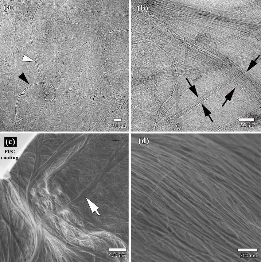 Figure 1: Cryo-EM micrographs of CNT/CSA solution, showing liquid crystalline phase development as a function of nanotube concentration (from (a) to (d) CNT concentration is increased). (a) Cryo-TEM image of an isotropic phase. Empty (black arrowhead) and filled with acid (white arrowhead) carbon nanotubes are recognized. (b) The beginning of aligned phase formation (black arrows) imaged by cryo-TEM. (c) Cryo-SEM of a network of aligned CNT domains (white arrow), coexisting with an isotropic phase. (d) Liquid crystalline domains at "dope" concentration for fiber spinning. Highly ordered, large (tens of microns long) liquid crystalline domains imaged by cryo-SEM.
