It is a long time that nature serves as a source of inspiration for the construction of useful materials and systems 1. Here, we present a simple, cost-effective and environmentally friendly bio-inspired approach for the creation of novel, hierarchical, porous drug polymorphs at nano and meso scales (Fig. 1). Our fabrication process utilizes the milk protein β-casein.
β-Casein is a 24 kDa amphiphilic and unstructured protein that possesses many properties facilitating its functionality in drug delivery systems, including strong self-association tendency into small core-shell micelles and excellent emulsification and stabilization abilities. We previously demonstrated the capability of β-casein micelles to efficiently encapsulate, stabilize in monomeric state, and deliver relatively high concentrations of hydrophobic drugs 2,3. Here, however, we suggest a new use of the drug-loaded protein carriers – as reactors for controlled creation of new semi-crystalline polymorphs (spherulites) of poorly water-soluble drugs. Our fabrication method involves first, encapsulation and stabilization of active agents within the hydrophobic core of the protein micelles. This is then followed by drug release and re-crystallization in the form of new polymorphs as a function of time, and in response to changes in environmental conditions. A proof of concept for the creation of the polymorphs is demonstrated with the desired drug celecoxib, a poorly water-soluble molecule used for the treatment of rheumatoid and osteo-arthritis, and a promising anticancer drug 4,5.
High-resolution scanning electron microscopy (HR-SEM) was our method of choice to study the particles structure and morphology at meso and nano scales. Cryogenic temperature transmission electron microscopy (cryo-TEM) 6 and light microscopy were complementary tools for the structural analysis, and x-ray diffraction (XRD) was employed to explore the drug physical state. These analyses revealed novel polycrystalline structures with a spherulitic, highly porous morphology. The high surface area, relatively small dimensions and monodispersity of such particles are highly desired properties in drug design that modern technologies seek for. Furthermore, tuning of the environmental conditions allowed a good control over the fine morphological characteristics of the drug particles. E.g., increasing the driving force for crystallization resulted in particles larger in size, and a gradual transformation from fine and highly arranged morphology into rough, open and somehow disordered morphology.
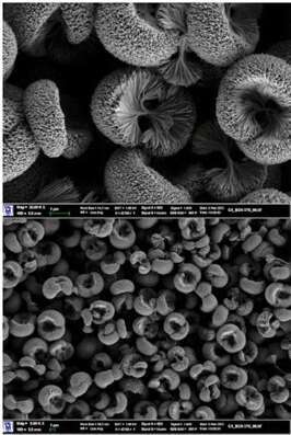
Figure 1. Drug polycrystals with uniform, porous and branched morphology.
References:
(1) Sarikaya, M.; Tamerler, C.; Jen, A. K.-Y.; Schulten, K.; Baneyx, F. Nat. Mater. 2003, 2 (9), 577–585.
(2) Bachar, M.; Mandelbaum, A.; Portnaya, I.; Perlstein, H.; Even-Chen, S.; Barenholz, Y.; Danino, D. J. Control. Release 2012, 160 (2), 164–171.
(3) Turovsky, T.; Khalfin, R.; Kababya, S.; Schmidt, A.; Barenholz, Y.; Danino, D. Langmuir 2015, 31 (26), 7183–7192.
(4) Vane, J. R.; Bakhle, Y. S.; Botting, R. M. Annu. Rev. Pharmacol. Toxicol. 1998, 38, 97–120.
(5) Thun, M. J.; Henley, S. J.; Patrono, C. JNCI J. Natl. Cancer Inst. 2002, 94 (4), 252–266.
(6) Danino, D. Curr. Opin. Colloid Interface Sci. 2012, 17 (6), 316–329.
