Since they were first introduced by Schnur and colleagues over 3 decades ago1, lipid nanotubes are being explored due to their principal roles in biology and medicine, as well as their potential in diverse applications. A specific class of synthetic lipids comprising diacetylene moieties in their tails, diacetylenic phospholipids, were shown to present distinct self-assembly, and form multilamellar nanotubes in alcohol-water solutions.
The first tube-forming amphiphilic lipid - 1,2-bis(10,12-tricosadiynoyl)-sn-glycero-3-phosphocholine designated DC8,9PC, have highly ordered and kinked alkyl chains which has been found to induce chiral self-assembly, regardless whether the constituent molecules are chiral2. Previous work focused on the properties and possible applications of the tubes, yet fundamental questions regarding their formation mechanisms were not addressed.
Using advanced electron microscopy techniques as the main research tools, we provide the first structural insight into the mechanism of formation of multilamellar DC8,9PC nanotubes. We show that these lipid nanotubes form through fast reorganization of the lipid membrane that involves unfolding, curling, bending and fusion. This new path provides an alternative pathway for the formation of peptide nanotubes, known to proceed from ribbon elements3, 4.
In addition to direct-imaging cryo-TEM we applied 3D cryo-TEM analysis using cryo-electron tomography (cryo-ET), a powerful method for 3-dimensional reconstruction of structures in solution5. Cryo-ET was applied for resolving overlapping morphologies and understanding complex membrane-tubular intermediate structures identified through the self-assembly pathway.
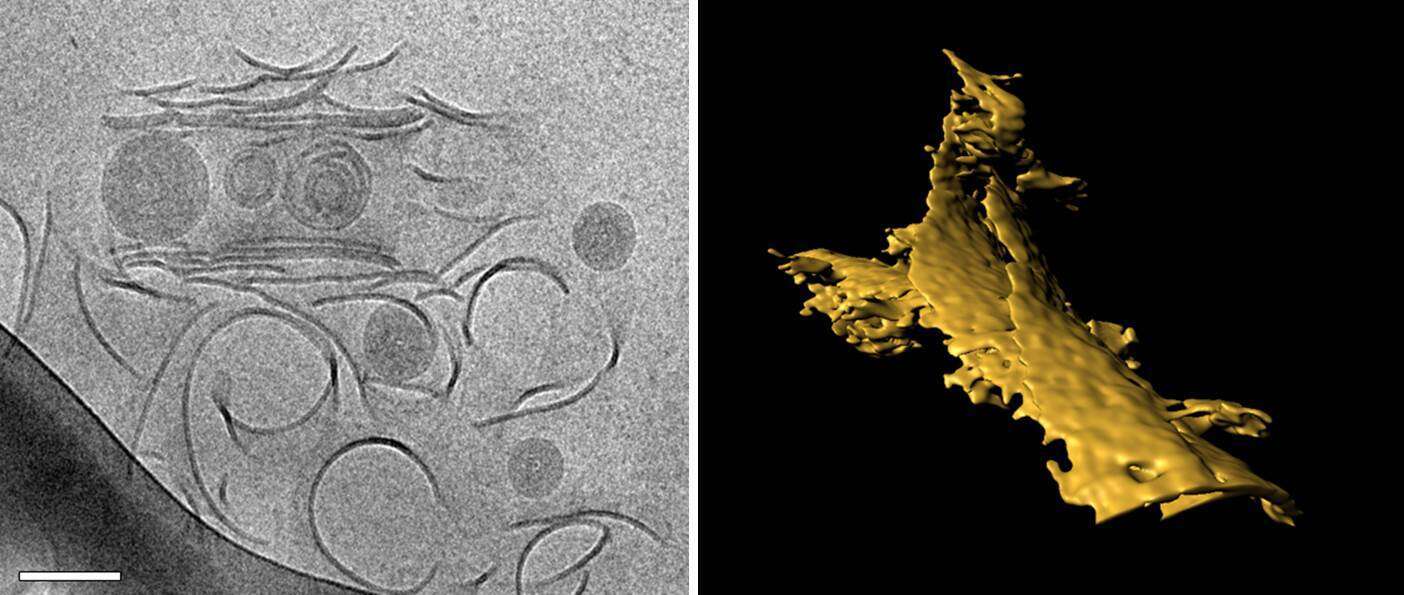
Fig 1. Left - Cryo-TEM image of DC8,9PC membranes in 70% EtOH/H2O solution captured during fast cooling to room temperature. Scale bar=200nm. Right - Segmented fragment from a tomogram showing membranes healing to form a multilamellar tubule wall.
References:
- Yager, P.; Schoen, P. E., Formation of Tubules by a Polymerizable Surfactant. Mol Cryst Liq Cryst 1984, 106 (3-4), 371-381.
- Pakhomov, S., et al., Chiral tubule self-assembly from an achiral diynoic lipid. Proc Natl Acad Sci U S A, 2003, 100(6): p. 3040-2.
- Ziserman, L.; Lee, H. Y.; Raghavan, S. R.; Mor, A.; Danino, D., Unraveling the mechanism of nanotube formation by chiral self-assembly of amphiphiles. J Am Chem Soc 2011, 133 (8), 2511-7.
- Ziserman, L.; Mor, A.; Harries, D.; Danino, D., Curvature instability in a chiral amphiphile self-assembly. Physical review letters 2011, 106 (23), 238105.
- Nudelman, F.; de With, G.; Sommerdijk, N.A.J.M., Cryo-electron tomography: 3-dimensional imaging of soft matter. Soft Matter 2011, 7(1), 17-14.
