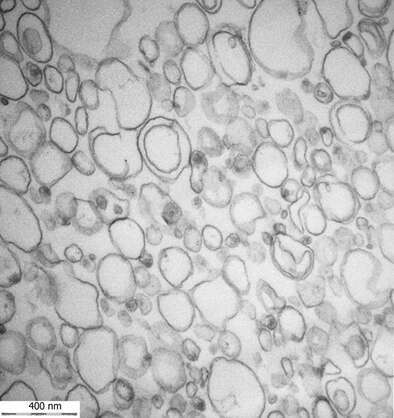
Extracellular vesicle (EV) is the common name of a heterogeneous group of lipid bilayer enclosed vesicles which can be released by practically all cells. These vesicles have been proved to have crucial roles in intercellular communication, signaling processes from inflammation to antigen presentation or tumor development and also in the transfer of genetic information. Especially the EV related biomarker and therapeutic applications made this research field attractive not only for basic but clinical research, too.
Although the story of extracellular vesicles started about 70 years ago we had to wait for the first transmission electron microscopic /TEM/ evidence of these mysterious little particles till 1981. Since then several great techniques and methods have been used or emerged to investigate and indentify EVs such as flow cytometry, atomic force microscopy, mass spectrometry, tunable resistive pulse sensing (TRPS), nanoparticle tracking analysis (NTA), microfluidic device etc., however, none of them is able to cover the whole size range of these vesicles /from 30nm till about 5µm/ as well as to demonstrate of their origin, the cells or tissue, moreover to distinguish the vesicular and non-vesicular particles. Not surpisingly, electron microscopy has become the golden standard of EV research, however it does not mean that TEM results would be generally accepted without any debate. The basic problem is the preparation of the extracellular vesicle populations. Numerous protocols have been applied by the different laboratories and the results published can be misleading. What can be accepted and which results should be refused, how can we avoid pitfalls during the vesicle preparation and whether the classical TEM can provide reliable results or instead everybody should use the fancy and expensive ultracryo microscopy?

Extracellular vesicles released by 5/4 cells

