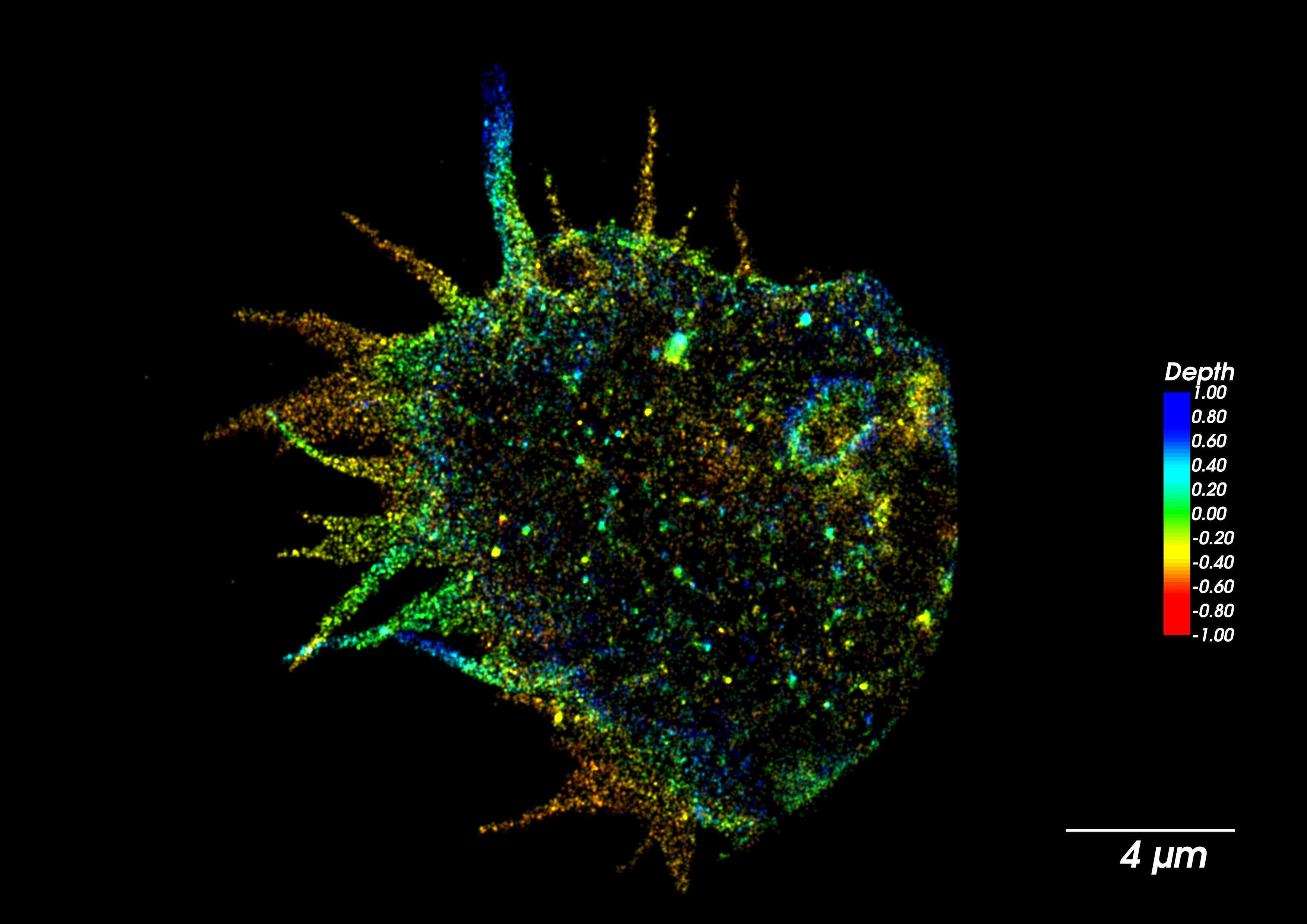
This image shows the 3D structure of the Amoeba’s pseudopodia being used for its motility and ingesting of nutrients. This image was taken using super resolution microscopy that allows sub-diffraction limit resolution of the actin filaments that form the pseudopodia unique and dynamic structure. Color gradient represents the depth of actin within the different pseudopodia protrusions at the tip of the Amoeba.
.