
Characterizing Mineral-Bearing Vesicles in Sea Urchin Embryos
2Department of Chemical Research Support, Weizmann Institute of Science, Rehovot
Sea urchin embryos have endoskeletons comprised of two calcitic spicules. Spicule growth takes place by the initial deposition of amorphous calcium carbonate (ACC)[1] in vesicles inside the spicule forming cells[2](PMCs). The calcium in the mineral bearing vesicles was recently reported to originate from body fluid internalization and in part from calcium channels[3]. Using cryo-scanning electron microscopy to image high pressure frozen and cryo-sectioned samples of embryos, we were able to detect several types of vesicles inside the PMCs. Some vesicles have granulated texture, some appear smooth, some have backscattered electrons signal, and some contain lipids or proteins. Performing EDS measurements under cryogenic conditions revealed that some of these vesicles are rich in sodium, while others give signals for potassium and calcium. We conclude that the compositional landscape of the vesicles in the PMCs is complex. Some of these vesicles fulfil a fundamental role in the mineralization of the spicules
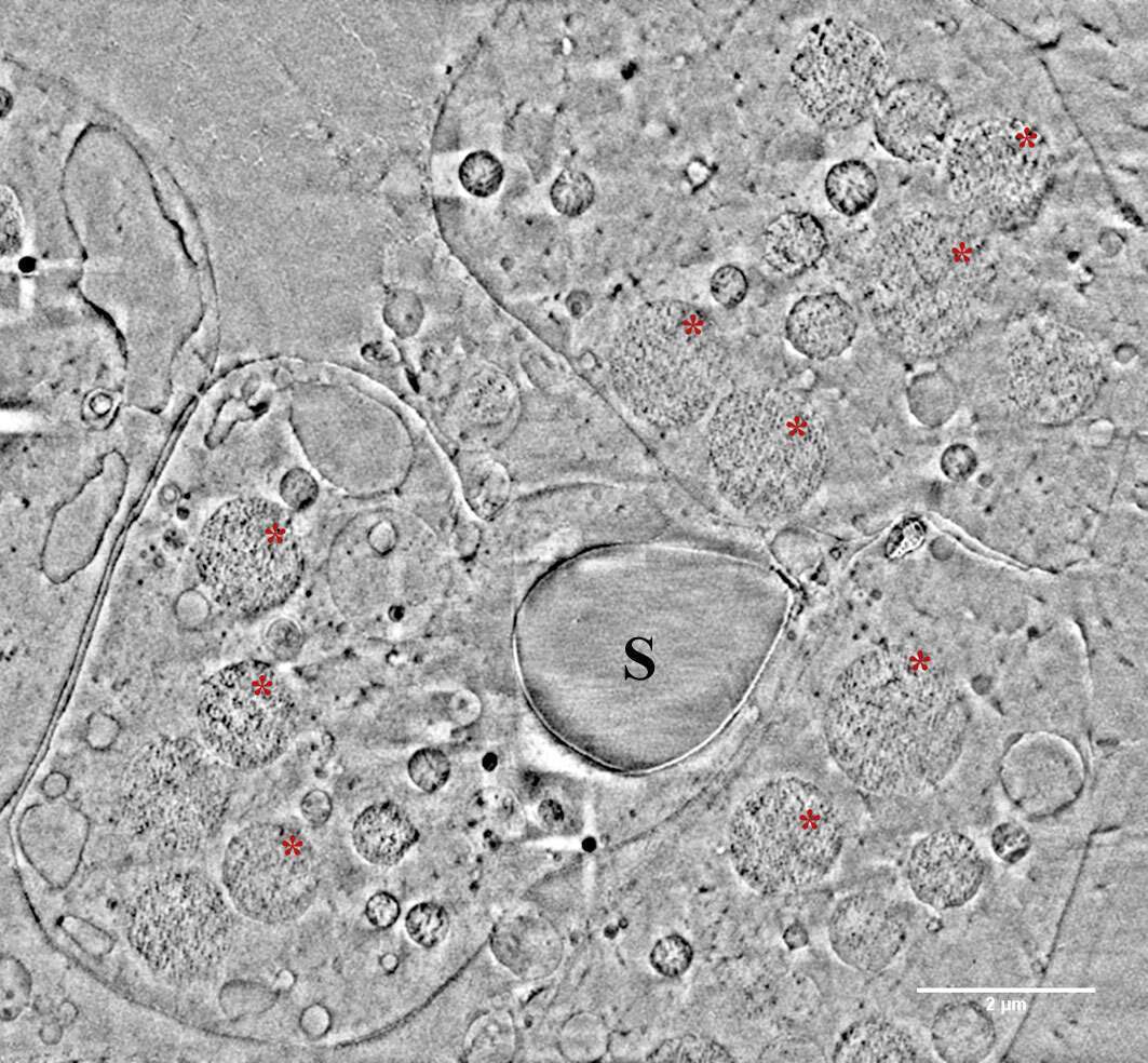
Figure 1 – Sea urchin embryo spicule (S) with its adjacent spicule forming cells. Red asterisks mark vesicles with granulated texture suspected to be ACC.
[1] E. Beniash, J. Aizenberg, L. Addadi, S. Weiner, P Roy Soc B-Biol Sci 1997, 264, 461-465.
[2] N. Vidavsky, S. Addadi, J. Mahamid, E. Shimoni, D. Ben-Ezra, M. Shpigel, S. Weiner, L. Addadi, P Natl Acad Sci USA 2014, 111, 39-44.
[3] N. Vidavsky, S. Addadi, A. Schertel, D. Ben-Ezra, M. Shpigel, L. Addadi, S. Weiner, Proc. Natl. Acad. Sci. U.S.A 2016, 201612017, 201610027-201618424.

Powered by Eventact EMS