
3d Compared to 2d Video Assistance Improves Early Outcomes in Minimally Invasive Mitral Valve Repair Surgery
2Cardiology, Sheba Medical Center, Tel Hashomer
3Anesthesiology, Sheba Medical Center, Tel Hashomer
Background:
Minimal invasive mitral valve surgery may reduce the need for blood products, lessen post-operative atrial fibrillation and decrease pain, compared to mid-sternotomy. However, these approaches are usually associated with longer cardiopulmonary bypass and cross-clamp times. The increasing use of new technology has facilitated the procedure and shortened its duration time. Mitral valve repair (MVr) is a reconstructive procedure and requires 3D vision.
Methods:
Of the 142 patients since 2004 who underwent minimally invasive MVr, the first 111 underwent the procedure through heart port (HP) video-assisted 2D technology, and the remaining 31 patients received HP video-assisted 3D technology. For the purpose of this study, we divided the patients into three groups: Group 1 – the first 50 patients (learning curve), Group 2 – the next 61 patients (2D technology), and Group 3 – 31 patients (3D technology).
Results:
Mean patient age was 50±14 years, and 103 (73%) patients were male.
Cardiopulmonary bypass and cross-clamp times were significantly shorter in Group 3 compared with Groups 2 and 1, respectively (110±20 vs. 124±23 vs. 139±27, p<0.001; and 79±25 vs. 86±18 vs. 97±18, p=0.001). There were no cases of in-hospital mortality, and none of the patients were converted to mid-sternotomy. Major post-operative complications included: revision due to bleeding or tamponade in 3 patients (2.1%), CVA in one patient (0.7%), and reversible renal failure in 3 patients (2.1%), with no significant differences between the groups. Early echo revealed that 96.7% of patients in Group 3, 90.2% in Group 2 and 89.8% in Group 1 had up to mild mitral regurgitation (MR) (p=0.171).
Conclusions:
By enhancing the dimension of surgical vision, minimally invasive MVr using 3D video-assisted technology provides significantly shorter cardiopulmonary bypass and cross-clamp times with low rates of residual MR. Improved technology will further facilitate the procedure and reduce its duration time.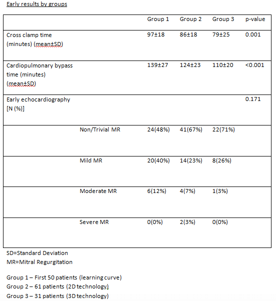
Powered by Eventact EMS