Prominent Differences in Cardiac Performance and Myocardial Properties between Right Ventricular and Left Ventricular-based Pacing Modes in Rats
2Regenerative Medicine & Stem Cell Research Center, Ben-Gurion University of the Negev
3Cardiology Department, Soroka University Medical Center
Background:
The biological effects of cardiac resynchronization therapy (CRT) are still elusive and require insights from animal models. However, CRT models currently relay on large mammals, which are expensive and not readily available. Recently, we introduced a device for long-term double-site epicardial pacing in rats, and found marked dyssynchrony during right ventricular pacing (RV) compared with both, left ventricular (LV) and Biventricular (BIV) pacing modes. Here we further characterized this model by hemodynamic recordings and analysis of myocardial properties following sustained tachypacing.
Objective:
To elucidate the effects of RV vs. LV-based pacing modes in rats.
Methods:
Electrodes were implanted either on the RA and RV or on the RV and LV. Wires were exteriorized through a back connector. Following recovery, rats were either used for admittance-based pressure-volume loop measurements or exposed to sustained RV vs. BIV tachypacing for 3 days. Western blotting was done conventionally.
Results:
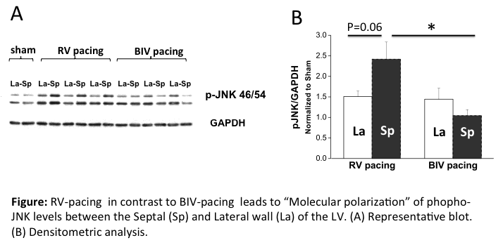
Sequential RA-RV pacing compromised systolic parameters including dP/dt max and stroke work, compared with RA pacing (n=8, p<0.001). In contrast, LV and BIV pacing modes markedly enhanced cardiac performance compared with RV pacing (n=8, p<0.001). Sustained RV tachypacing (n=6), but not BIV tachypacing (n=6), increased the QT interval by 6.63±3.1 ms relative to baseline and dispersed refractoriness between the right and left pacing sites (10.0±3.8ms, p<0.05). In addition, increased expression of Osteopontin and activation of CAMKII and JNK signaling (Figure) were found in the early-activated septal tissue of RV-paced group compared with the BIV-paced group.
Conclusion: Our data support the notion that this is an attractive new model to study the biological consequences of dyssynchrony and CRT. The new biochemical findings can shade light on fundamental effects of the different pacing modes on the tissue level.

Powered by Eventact EMS