
Validation of and Comparison between Methods to Assess Diastolic Function by Cardiac CT
2Radiology, Rambam Medical Centre
3Medicine, Technion-Israel Institute of Technology
Background:
Noninvasive assessment of left ventricular (LV) diastolic function is typically performed using echocardiography however interpretation of results is complex and imperfect. Recently two methods for evaluating diastolic function using cardiac computed tomography (CT) have been published, the first based on echocardiographic parameters (echo-based method) and the second utilizing left heart volume changes (volume method), however the methods have not been validated on sequential patient studies and no comparison has been made between the methods. We aimed to compare these methods in a consecutive group of patients using echo as the gold standard.
Methods:
100 consecutive patients who underwent cardiac CT using helical scanning and who had a full echo examination within 2 months were studied. 22 patients were excluded due to atrial fibrillation, mitral mechanical prostheses, or significant mitral valve disease. The remaining 78 patients were classified as normal, indeterminate or grades 1-3 diastolic dysfunction based on ASE echo guidelines. CT analysis involved calculating the early diastolic mitral flow (Eflow) and tissue velocity (e`) (echo-based method) and left atrial (LA) volumes and function (volumetric method).
Results:
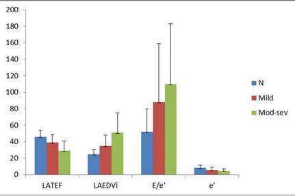
By echocardiography, 21 patients had normal diastolic function, 43 abnormal and 13 indeterminate results. Results are summarised in the chart below. By ROC analysis, the optimal parameters by echo-based method were Eflow/e` with area under the curve (AUC) 0.79 [0.67-0.92] and e` with AUC 0.78 [0.65-0.91] and by the volume method, LA end-diastolic volume (LAEDV) with AUC 0.85 [0.76-0.94] and LA emptying fraction (LATEF) with AUC 0.83 [0.74-0.93].
Conclusion:
Both CT methods can differentiate between normal and abnormal diastolic function relative to echocardiography as a reference. The volume method appears to be more accurate than the echo-based technique.
Powered by Eventact EMS