Establishment of the Vascular Injury Model of Retinopathy of Prematurity in vitro
Background: The early stage of retinopathy of prematurity (ROP) mainly presented as retinal vascular retardation. The most important causes of ROP are hyperoxia/hypoxia injury, inflammation, etc. ROP rat model, namely the oxygen-induced retinopathy (OIR) model, was conducted by intermittent hyperoxia and hypoxia stimulation for 14 days in neonatal rats. During 0-14 days, retinal vasculature appeared dysplasia, and failed to reach the edge of the retina. Although the animal model is mature, the cell model which is consistent with the vascular endothelial cell injury of ROP is lacked.
Objective: This study was designed to explore the in vitro model of retinopathy of prematurity.
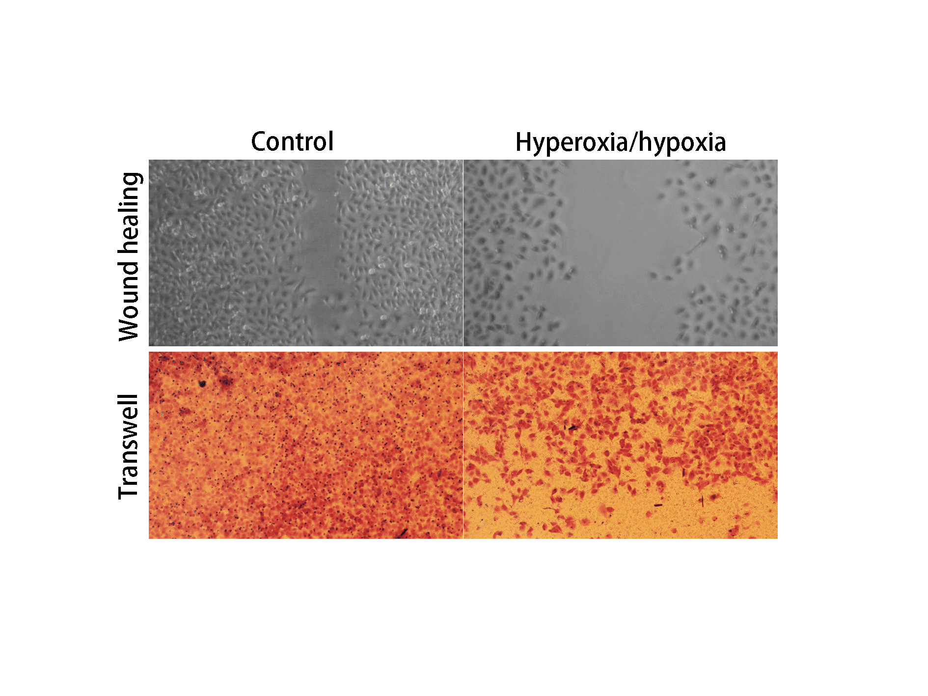
Methods: The experiment group was treated with hyperoxia-induced complete medium (containing 50μmol /L H2O2 solution) for 15 minutes and hypoxia-induced complete medium (containing 50μmol /L CoCl2 solution) for 4 hours. Two cycles of hyperoxia/hypoxia stimulation were conducted. Wound healing kinetic and transwell migration assay were used to test the proliferation and migration abilities of the HUVEC.
Results: After two cycles of stimulation, cells in the experiment group were smaller than those in the control group. In vitro wound healing kinetic showed that the hyperoxia/hypoxia stimulation compromised HUVEC wound closure; transwell migration assay confirmed that migrated cells were more in the control group, indicating truncated ability of migration in the experiment group.
Conclusion: The proliferation and migration abilities of endothelial cells are damaged in the experiment group, which are consistent with the inhibition of angiogenesis of ROP in early stage. Therefore, in light of the rat OIR model, the cycled hyperoxia/hypoxia cell model has potential in mimicking ROP vascular injury.

Powered by Eventact EMS