
Differentiating between Cancer and Inflammation: a Novel Metabolic-Based Method for Functional CT Imaging
2Cyclotron-Radiochemistry-MicroPET Unit, Hadassah-The Hebrew University of Jerusalem, Jerusalem, Israel
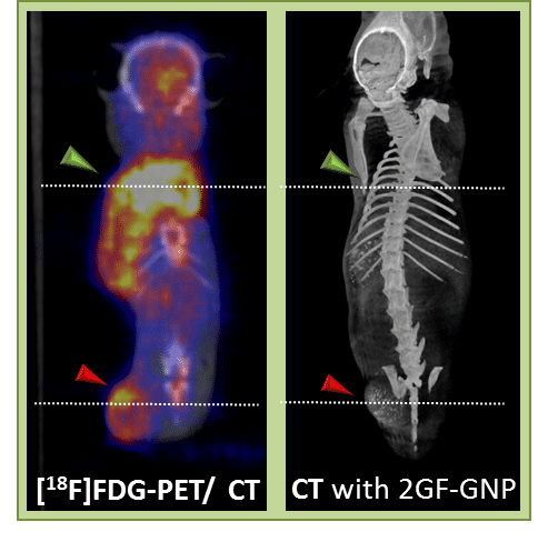
One of the main limitations of the highly used cancer imaging technique, PET-CT, is its inability to distinguish between cancerous lesions and post treatment inflammatory conditions. The reason for this lack of specificity is that [18F]FDG-PET is based on increased glucose metabolic activity, which characterizes both cancerous tissues and inflammatory cells. To overcome this limitation, we developed a nanoparticle-based approach, utilizing glucose-functionalized gold nanoparticles as a metabolically targeted CT contrast agent. Based on previous knowledge that newly formed blood vessels in growing tumors differ from those in inflammation, we hypothesized that the proposed technique may provide the ability to differentiate tumors from non-malignant metabolically active processes. Indeed, our approach has demonstrated, by both CT and quantitative measurements, specific tumor targeting and distinction between cancer and inflammation, in a combined tumor-inflammation mouse model. By contrast, FDG-PET/CT scans showed no visible nor quantitative differentiation between the two. In conclusion, our new concept of functional CT imaging overcomes the main drawbacks of the currently used FDG-PET and provides a new set of capabilities in cancer detection, staging and follow-up. This novel technology can be applicable to a wide range of cancers which exhibit high metabolic profiles.
Powered by Eventact EMS