
Benefits of Pressure Actuation for Flow Control in Microfluidics
2., Micronit, Enschede, Netherlands
3RIKILT, Wageningen University and Research, Wageningen, Netherlands
Abnormal cell phenotype, gene expression and metabolic activity of cells cultured in vitro compared to in vivo have questioned the utility of traditional cell culture methods. Due to this issues a new generation of in vitro models (organ on chip) has emerged which recreates to some extent, the microenvironment of living cells to preserve their physiological activity [1]. In this context, Fluigent and Micronit have developed a versatile fully integrated organ on a chip platform which recreates the main biochemical and biophysical features of the native environment of cells in order to induce their growth and differentiation as functional tissues.
The platform comprises a flow controller connected to a resealable glass chip separated into two flow chambers by a transversal porous membrane (Fig1). To reproduce the in vivo spatial partition of cells, different cell types can be seeded on each side of the membrane and perfused with their specific medium (Fig 2). Thanks to the great versatility of the flow controller, biochemical gradient, air-liquid and liquid-liquid interfaces can be implemented in the chip to replicate the biochemical environment of living cells. Finally, specific physiological mechanical constraints like cyclic tissue stretching (muscle, lung…) or flow induced shear stress (laminar or pulsatile) can also be faithfully recreated by the platform.
[1] Verhulsel M et al, A review of microfabrication and hydrogel engineering for micro-organs on chips. Biomaterials. 2014; 35(6):1816-32
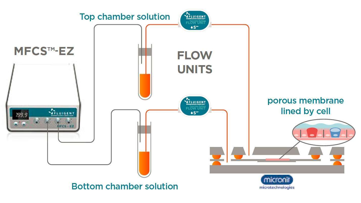
Figure 1: Scheme of the platform: flow control system connected to microfluidic chip
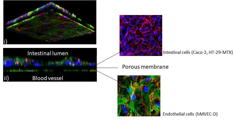
Figure 2 : Gut-on-chip model : coculture of intestinal and endothelial cells.(i) Basal and (ii) transversal view of the membrane. (Nuclei: blue, actin: green and tight junctions: red), pictures from Dr Meike van der Zande, RIKILT
Powered by Eventact EMS