
Investigation of Cystoliths in an Aerating Roots of Orchidaceae
2Institute of Chemistry, The Hebrew University of Jerusalem (retired), Jerusalem, Israel
The functions ascribed to calcium in living plant cells are well known. It has been established that a decrease in the inhibition of cytoplasmic colloids by adsorbed Ca2+ produces reduced permeability; while the binding of Ca2+ ions to pectins of the cell wall increases wall rigidity. The absolute amounts of calcium required are very low.1 Biogenic amorphous materials are prominent in the field of biomineralization. For example, amorphous calcium carbonate has been shown to be a precursor to calcite.2 Plant cystoliths are structures, mineralized with calcium carbonate, that are formed by specialized cells in the leaves of certain plants.3
Here we report on the observation of calcium carbonate microparticles in aerating roots of Orchidaceae. We observed numerous microparticles and their bundles in SEM (scanning electron microscope) images acquired from freshly prepared cross-sections of an aerating root. According to EDS (energy dispersive X-Ray spectroscopy), the microparticles are mainly composed of Ca, C and O, thereby supporting identification of calcium carbonate (containing minor amounts of K). Most particles have either needle - or plate like morphology. They are distributed over the entire root cross section. The location and morphology of the microparticles indicates that solidification/crystallization of calcium carbonate occurred inside the root cells. To the best of our knowledge this is the first observation of a calcium carbonate cystholith in an aerating root of Orchidaceae.
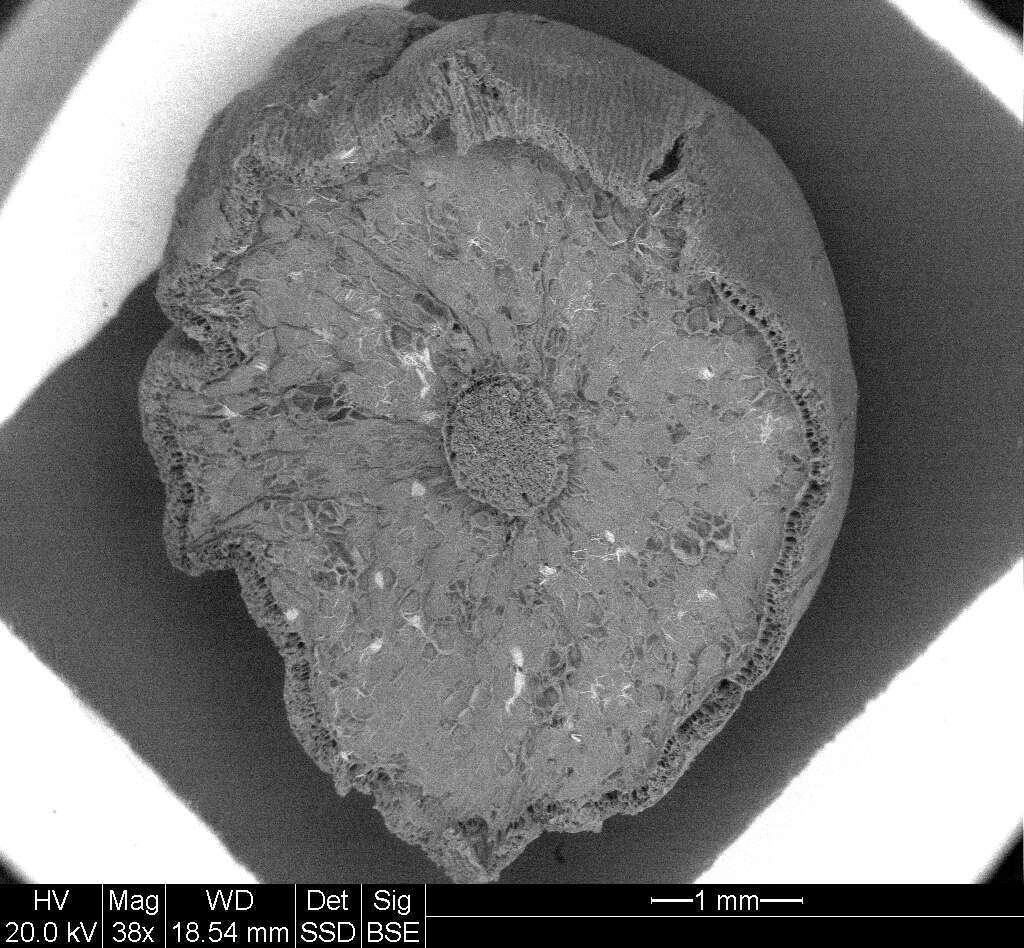
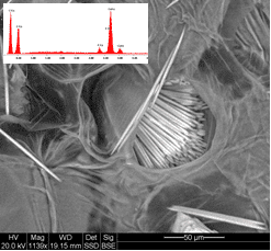
SEM images of a root cross section. Low resolution image of a root cross section with back scattered electrons (left image). In a higher magnification image (right image) with back scattered electrons, needle-like crystallites are clearly visible. The insert contains the EDS spectrum acquired from a bundle of needles in which the characteristic peaks of C, O, Ca and K are clearly visible (right image).
References: (1) Burstrֳom, H.G. Biol. Rev. (1968), 43, pp. 287-316. (2) Beniash, E. et al. Proc. R. Soc. Lond. B, (1997), 264, 461-465. (3) Gal.A. et al. A European Journal, (2012), 18, 10262-10270.
Powered by Eventact EMS