
The Role of Left Ventricular Circumferential Rotation in Functional Mitral Regurgitation Mechanism
Background: Functional Mitral Regurgitation (FMR) is caused by left ventricular (LV) dysfunction. Past research has described the circumferential components of LV contraction including twist, but their role in FMR remains unclear. We hypothesized that LV systolic twist would be affected in patients with FMR and therefore evaluated the relationship between LV twist and FMR utilizing novel cardiac Magnetic Resonance Imaging (MRI) image processing techniques.
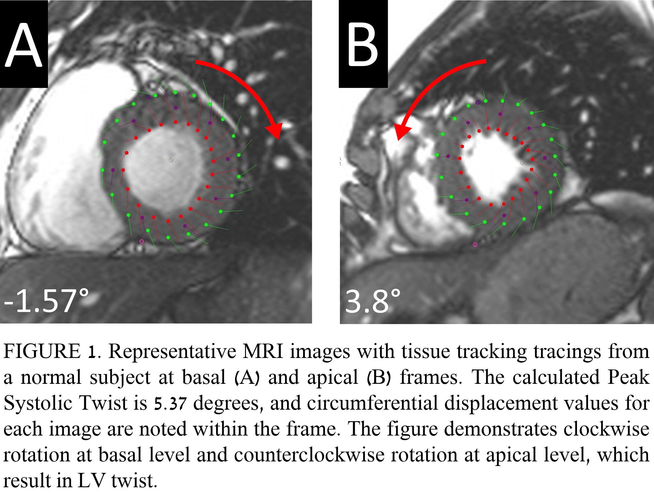
Materials and Methods: Normal subjects (n=40), low EF (EF<50%) patients without FMR (n=17), and low EF patients with FMR (regurgitant volume (RVol)>10 mL; n=20) were studied. All subjects underwent MRI for clinical indications. 2D tissue-tracking analyses were conducted based on short axis and long axis images using Circle CVI42 version 5.3.0.821 software. Peak Systolic Twist was calculated as the difference between Peak Apical Circumferential Displacement and Peak Basal Circumferential Displacement (figure 1). RVol was calculated as the difference between stroke volume and the forward flow in the ascending aorta.
Results: Peak Systolic Twist was significantly decreased in FMR patients (Median: 1.1 deg, IQR: 0.6-2.53 deg) as compared to normal subjects (Median: 2.25 deg, IQR: 1.6-3.02 deg; p=0.02), while no significant difference was observed when comparing normal subjects to low EF patients (Median: 2.35 deg, IQR: 0.93-3.8 deg, p=1.00). RVol negatively correlated with Peak Systolic Twist (r=-0.4; p<0.001) and Peak Apical Circumferential Displacement (r=-0.33; p<0.01), and positively correlated with Time to Peak Apical Circumferential Displacement (r=0.34; p<0.01).
Conclusion: Peak Systolic Twist is significantly decreased in patients with FMR and is correlated, along with several other circumferential components of LV contraction, with its severity, potentially adding to the understanding of FMR pathogenesis.

Powered by Eventact EMS