
Ultrastructural Imaging of Salmonella-Host Interactions Using Super Resolution Correlative-Light-Electron Microscopy of Bioorthogonal Pathogens
2Nanoscopy for Nanomedicine, Institute of Bioengineering of Catalonia, Barcelona, Spain
3Department of Electron Microscopy, Leiden University Medical Center, Leiden, The Netherlands
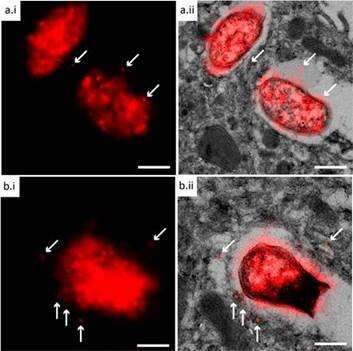
Bioorthogonal chemistry has been a major breakthrough technique in the study the interaction of this host-pathogen interaction. Through hijacking of the cell wall biosynthetic machinery with biorthogonal analogues of cell wall components, the cell walls of intracellular pathogens have been visualized selectively within host cell phagosomes. We recently reported an approach that allows the visualization of bacteria within the ultrastructural context of the host cells by imaging bioorthogonal bacteria using correlative light and electron microscopy (CLEM). After sectioning frozen hydrated cells down to a thickness of 80 nm followed by an on-section copper-catalyzed Huisgen cycloaddition reaction (ccHc) and subsequent EM-imaging, we could image the location of bioorthogonal groups within the structure of a cell.
However, one major limitation to date has been the resolution of the confocal imaging: whereas the resolution of the EM micrograph is in the order of 1 nm, that of the fluorescence is limited by the van Abbe diffraction limit of half the wavelength of the photon (approx. 250 nm). To circumvent this, we have developed an improved approach: by using stochastic optical resolution microscopy (STORM) on the sections, to bring the resolution of the fluorescent signal in closer alignment with that of the TEM we can now image bioorthogonally labelled pathogens inside phagocytes with optical resolutions of ~20nm, yet with full ultrastructural information on where in the cell the pathogen resides.
References:
van Elsland, D. M.; Bos, E.; de Boer, W.; Overkleeft, H. S.; Koster, A. J.; van Kasteren, S. I. Detection of bioorthogonal groups by correlative light and electron microscopy allows imaging of degraded bacteria in phagocytes. Chemical Science 2016, 7 (1), 752.
Van Elsland, D. M.; Bos, E.; Pawlak, J. B.; Overkleeft, H. S.; Koster, A. J.; Van Kasteren, S. I. Correlative light and electron microscopy reveals discrepancy between gold and fluorescence labelling. Journal of Microscopy 2017
Powered by Eventact EMS