The development of four-dimensional ultrafast electron microscopes (4D UEM) breaks the time resolution limit of conventional transmission electron microscopy techniques and enables sample imaging with fs temporal and nanometer spatial resolutions [1]. This promising capability is based on photon-electron interactions mediated by the evanescent field localized around the imaged nanostructure [2-4] and is known as photon-induced near-field electron microscopy (PINEM) [1-3]. Despite many reports on ultrafast dynamics of plasmons and phonons [5-6], the full potential of imaging via the interactions between electrons and the light field is not completely understood yet. In this work, we developed the PINEM technique for novel imaging capabilities in two new scenarios: Scanning resonant near-field electron microscopy and Semi-infinite far-field electron microscopy.
In scanning resonant near-field electron microscopy [7] many nanostructures have spectral responses that are highly sensitive to the excitation frequency. To enable coupling into such resonant modes with PINEM, a tunable wavelength laser for excitation is required. Here we demonstrate this technique with a silver nanowire by employing an optical parametric amplifier as a wavelength-tunable light source. The plasmonic mode energy was resolved down to the laser linewidth; 20 meV in this work. This scanning resonant electron microscopy not only reveals the ultrafast dynamics of a nanostructure, but can also determine the oscillatory energy with meV resolution. Moreover, it becomes possible to directly image optically sensitive low-energy modes such as phonons and Raman vibrations which are too weak to be observed by conventional PINEM and EELS.
In semi-infinite far-field electron microscopy [8] we investigated electron-photon interactions in the semi-infinite vacuum far-field rather than using the evanescent near-field of a nanostructure as the medium for photon-electron energy exchange as in PINEM. We observed the depletion of the zero-loss peak (ZLP) in EELS and the broadening of the electron energy distribution as the tilt angle increased. Remarkably, this implies that almost all electrons interacted with the photon field. This strong coupling in the far-field greatly extends the reach of PINEM by increasing the overall photon-electron interaction cross-section and thus producing a significantly enhanced signal. This work implies a potential imaging technique assisted by refracting, absorbing, or reflecting interfaces.
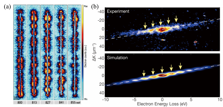
Fig. 1 New imaging capabilities through the PINEM technique in two new scenarios: (a) Imaging plasmonic standing waves by a tunable excitation in the UEM (b) Observation and simulation of the energy and momentum exchanges between electron and photon in UEM diffraction mode.
References
[1] A. Zewail, “Four-Dimensional Electron Microscopy”, Science 328, 187-193. (2010)
[2] B. Barwick, D. Flannigan and A. Zewail, "Photon-induced near-field electron microscopy". Nature 462, 902-906. (2009)
[3] S. T. Park, et. al., "Photon-induced near-field electron microscopy (PINEM): theoretical and experimental." New J. Phys. 12, 123028 (2010).
[4] F. de Abajo. “Optical excitations in electron microscopy.” Rev. Mod. Phys. 82, 209. (2010)
[5] D. Cremons, et. al., "Defect-mediated phonon dynamics in TaS2 and WSe2." Struct. Dyn. 4, 044019. (2017)
[6] L. Piazza, et. al., “Simultaneous observation of the quantization and the interference pattern of a plasmonic near-field.” Nat. Commun. 6, 6407. (2015)
[7] E. Pomarico, et. al., “meV resolution in laser-assisted energy-filtered transmission electron microscopy”, ACS Photonics. (2017)
[8] GM Vanacore et. al., “From attosecond to zeptosecond coherent control of free-electron wave functions using semi-infinite light fields”. arXiv:1712.08441

