Investigation of the Spread Behavior of Plant Roots Mucilage in Sandy Soil using X-Ray Computed Tomography
Root mucilage plays an important role for rhizosphere hydraulic properties and hence, for soil-plant water relations. There are a number of studies addressing the chemical composition of root mucilage and its different properties. However, there is hardly any information on the spatial distribution of mucilage. The main objective of this study was to develop a method for visualization of mucilage in situ based on its specific properties. As visualization tool X-ray CT was used. This technique enables 3D visualization of roots and soil matrix.
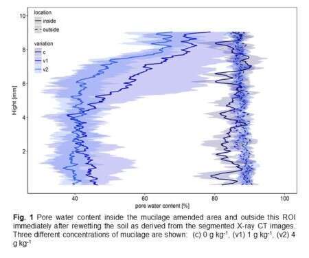
Two experimental approaches were used to localize mucilage, both based on the fact that mucilage turns hydrophobic upon drying. First a layer of mucilage amended quartz sand (0, 1, 4 g kg-1) was placed within a sand column. Columns were saturated from the bottom and scanned immediately, one and seven days after saturation. Within a region of interest (ROI) the volume fractions of water and air filled pores were segmented for all time steps (Fig. 1). In the area where the mucilage amended soil had been placed, air filled porosity was much higher than in the control treatment (0 g kg-1). No differences could be detected between 1 and 4 g kg-1. Air filled porosity decreased slowly with time indicating an increase of wettability.
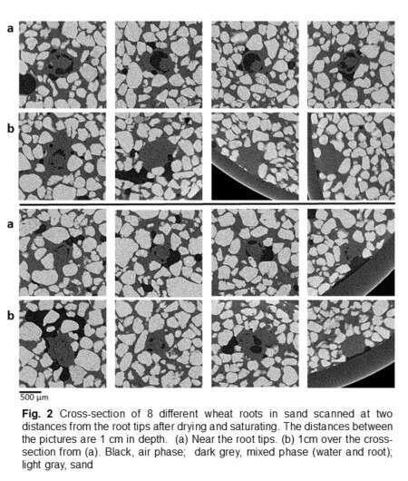
The second approach was an in-situ experiment with wheat roots growing into wet sand. After four days the system was cooled and dried out. Afterwards, the sand was saturated and the samples were scanned at two distances from the root tips. Images show air filled pores at the root-soil interface. Unfortunately it is not possible to differentiate between loss of soil-root contact as a result of shrinking and swelling and air filled pores introduced by mucilage induced hydrophobicity (Fig. 2).


Powered by Eventact EMS