
FLUORESCENT MOLECULARLY IMPRINTED POLYMER-BASED CORE/SHELL/SHELL NANOPARTICLES FOR STAINING SIALIC ACID RESIDUES ON TUMOR CELLS
2Department of Biomedical Sciences, Faculty of Health and Society, Malmö University
Cancer is a leading cause of death worldwide, and its early detection and resultant treatment contributes significantly to patient recovery and survival. Detection is currently based on magnetic resonance imaging and computed tomography, methods that are expensive, while processing of the results is time consuming1. There is a need for low-cost cancer-detection techniques that give conclusive results in the shortest time possible. Molecularly imprinted polymers (MIPs) targeting tumor markers on cancerous cells may provide a cheaper solution for cancer detection. Thin MIP layers immobilized on particle platforms are known to give faster response times and increased selectivity in comparison to bulk MIPs. It has been reported that a fluorescent monomer can be incorporated into the MIP layer2-3, allowing for faster detection of the target group, thus significantly shortening the turn-around time for biopsies.
Changes in sialylation patterns of cell surface glycoproteins indicate malignancy4. Here, we present the development of MIPs that target sialic acid-terminated glycoproteins (SA MIPs), prepared as a thin layer on a polystyrene core/silica shell nanoparticle platform. A fluorescent monomer is incorporated into the MIP layer, and upon binding of the target group to the specific binding pockets in the MIP, the fluorescence signal is enhanced. Transmission electron microscopy (TEM) and scanning electron microscopy (SEM) are used for structural characterization. To validate the specificity, fluorescence changes of MIPs in the presence and absence of template are compared to their corresponding non-imprinted polymer particles (NIP). Initial binding experiments with tumor cells using fluorescence microscopy demonstrate that the presented technique shows promise as a cheaper alternative to current detection methods, while allowing for relatively shorter analysis of biopsy results.
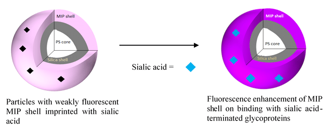
Figure 1: Detection of sialic acid-terminated glycoproteins by fluorescent core/shell/shell MIP particles
References
[1] Kakushadze, Z.; Raghubanshi, R.; Yu, W., (2017) Estimating Cost Savings from Early Cancer Diagnosis. Data 2: 30-45
[2] Shinde, S.; El-Schich, Z.; Malakpour, A.; Wan, W.; Dizeyi, N.; Mohammadi, R.; Rurack, K.; Gjörloff Wingren, A.; Sellergren, B., (2015) Sialic Acid-Imprinted Fluorescent Core–Shell Particles for Selective Labeling of Cell Surface Glycans. Journal of the American Chemical Society 137: 13908-13912
[3] Wan, W.; Descalzo, A. B.; Shinde, S.; Weißhoff, H.; Orellana, G.; Sellergren, B.; Rurack, K., (2017) Ratiometric Fluorescence Detection of Phosphorylated Amino Acids Through Excited-State Proton Transfer by Using Molecularly Imprinted Polymer (MIP) Recognition Nanolayers. Chemistry – A European Journal 23: 15974-15983
[4] Dube, D. H.; Bertozzi, C. R., (2005) Glycans in cancer and inflammation — potential for therapeutics and diagnostics. Nature Reviews Drug Discovery 4: 477-488

Powered by Eventact EMS