High-Resolution Episcopic Microscopy (HREM, developed by Tim Mohun, CR-UK) is a technique for visualising samples, such as embryos, plants and small organs. Practically, sample size can vary between 200µm3 to 1.5cm3. HREM generates a large stack of successive 2D sectioned images, as thin as 1 μm. The resolution of the images approaches that of conventional histology and since they are perfectly aligned, they can be used to construct very detailed 3D models. HREM images typically have voxel resolutions of 1-8 μm3. At this resolution it is possible to identify features such as individual nuclei and cells, nerves and blood vessels, which may not be detectable using lower-resolution techniques such as optical projection tomography (OPT), micro-magnetic resonance imaging (μMRI) or micro-computed tomography (μCT).
HREM data can be analysed either as 2D image stacks or by 3D rendering. Building detailed 3D models from images of individual sections has always proved difficult, since accurate alignment of the images is required. In addition, the sections themselves often become distorted as they are cut, captured and stained. Using HREM avoids these problems by sequentially imaging the face of the block, rather than imaging individual cut sections.
Using a multi-channel HREM, various features can be simultaneously visualized on top of morphology. This includes histological staining and gene expression patterns that can be segmented and quantified in 3D. The commercial version of the HREM was introduced only two years ago and it is currently used in handful laboratories worldwide. The multi-channel HREM at the Volcani Center is used for embryology, plant, medicine and agricultural studies, as well as for developing new capabilities of the HREM system itself.
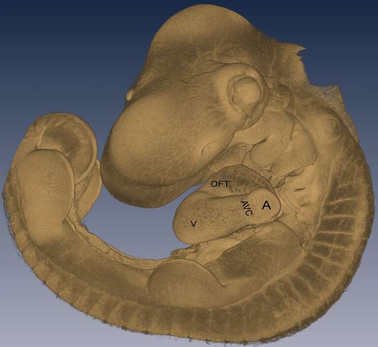
Using the HREM system we modeled the chicken embryo (blastoderm) in high 3D resolution which allowed us to study the earliest embryonic stages.
The chick blastoderm is composed of a circular epithelial disk termed the Area Pellucida, surrounded by a multi-layered ring called the Area Opaca. Within the first 12 h of incubation the embryo undergoes blastulation, during which a second layer - the hypoblast is formed. These stages are known as Eyal-Giladi and Kochav stages X-XIII EG&K. In stages XI-XIII EG&K, The hypoblast progressively formed anteriorly.
Following oviposition, blastoderms can undergo diapause that is characterized by a reversible suspension of metabolism and developmental. We characterized blastoderms challenged their ability to diapause for prolonged time under different temperature conditions. Using the HREM, we found that at oviposition, the predominant developmental stages of embryos from young flocks corresponds to XI. Following prolonged diapause, of up to 28 days, we found that while at 18oC the developmental stage progresses, at 12oC beyond 7 days, it stops. Further, we analyzed the effects of storage on the cytoarchitecture, mitotic index, cell death and embryonic survival until hatching. We show that at 18oC the embryos undergo dramatic cellular remodeling, manifested by maladaptive recesses, increased apoptosis and substantiate embryonic mortality. In contrast, storage at 12oC is advantageous in terms of developmental progression, cytoarchitecture maintenance, apoptosis rate and embryonic vitality.