Sea urchin larvae have endoskeletons comprised of two calcitic spicules. Large amounts of calcium must assemble in the larva to build the spicules, requiring substantial calcium uptake from the water or the food into spicule-forming cells (PMCs). PMCs uptake seawater through endocytosis1 into a complex network of vacuoles. Within the PMCs, calcium ions are translocated from the seawater vacuoles to various organelles and vesicles where they accumulate, and subsequently precipitate as a mineral2. The amorphous precipitates are finally translocated to the spicule, where they crystallize.
Here we address the question of the form in which calcium ions are stored in different locations in the cell –dissolved, solid and if so what is the solid phase? In order to locate and characterize calcium content in individual vesicles we apply cryo-soft X-ray microscopy on PMCs. The presence of concentrated calcium ions was detected by imaging the cells in the energy range before and after the calcium L-absorption edge (Figure 1). We characterized the chemical environment of the calcium ions using X-ray absorption spectroscopy and determined that some of these particles are composed of different forms of highly disordered calcium carbonate. We also developed a method for quantitative measurement of ion concentrations by EDS, whereby we follow the development of vesicle content. These data shed light on the intracellular transport and concentration pathways of calcium ions, eventually leading to a deeper understanding of mineral formation.
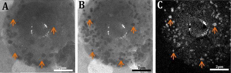
Figure 1 - (A) A spicule-forming cell imaged at 342eV, below the Ca L-absorption edge. (B) The same cell imaged on the Ca-absorption edge at 352.1eV. Many particles appear to absorb at this energy (dark particles) – corresponding to areas containing concentrated Ca. (C) “Ca-map” of the same cell. The map was obtained by subtracting the image in (A) from the image in (B). Representative Ca-containing particles are marked by arrows. The varying intensities of these particles are indicative of varying Ca concentrations
References
- Vidavsky, S. Addadi, A. Schertel, D. Ben-Ezra, M. Shpigel, L. Addadi, S. Weiner, Calcium transport into the cells of the sea urchin larva in relation to spicule formation. Proc. Natl. Acad. Sci. U.S.A, 113(45), 12637-12642,2016.
- Beniash, J. Aizenberg, L. Addadi, S. Weiner, Amorphous calcium carbonate transforms into calcite during sea urchin larval spicule growth. P. Roy. Soc. B-Biol. Sci., 264 (1380), 461-465, (1997).

