Vertebrate bone is a specialized connective tissue (1) with a highly organized hierarchical structure (2). Bone consists of carbonated hydroxyapatite nanocrystals formed within an organic matrix of collagenous fibers and various biological molecules. The elongation of long bones occurs through a mechanism of endochondral ossification which happens exclusively at the growth plate (3). In the endochondral ossification pathway mesenchymal cells differentiate to produce intermediate cartilage tissue, which is later replaced by bone tissue. The growth plate is also a relevant region for bone disorders, infections and a target area for secondary metastases during breast cancer. To date, a high-resolution characterization of the microstructure of the growth plate under near-physiological conditions is missing. This research aims to improve our understanding of both the growth plate structure and endochondral bone formation pathways. In order to explore this clinically crucial region of bone biology, we have avoided the standard freeze-fracture technique used in cryo-SEM, and instead have developed an innovative approach to examine fully hydrated mice tibiae growth plates under cryogenic conditions, using cryo-planing (Figure1). The technique consists of rapid freezing of the tissue after it was infiltrated with a cryo-protectant (glycerol) and then cryo-planing followed by block-face imaging. This technique allows visualization of both the growth plate chondrocytes (GPC) and the mineralization front (MF) up to the resolution of a few nanometers. Integration of these images with fully hydrated microCT analysis will, in the future, provide a micrometer-scale 3D characterization of both the non-mineralized soft-tissue and the mineralized components of the hard tissue.
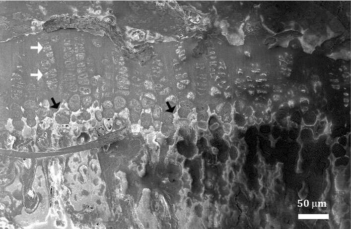
Figure 1. Cryo-SEM block-face micrograph of a mouse tibia showing the growth plate chondrocytes (white arrows) and the mineralization front (black arrows).
References:
- Bourne GH (1957) The Biochemistry and Physiology of Bone (Elsevier).
- Weiner S & Traub W (1992) Bone structure: from angstroms to microns. The FASEB Journal 6(3):879-885.
- Irving J (2012) Calcium and phosphorus metabolism (Elsevier).

