Structural Biology, Weizmann Institute of Science, Rehovot
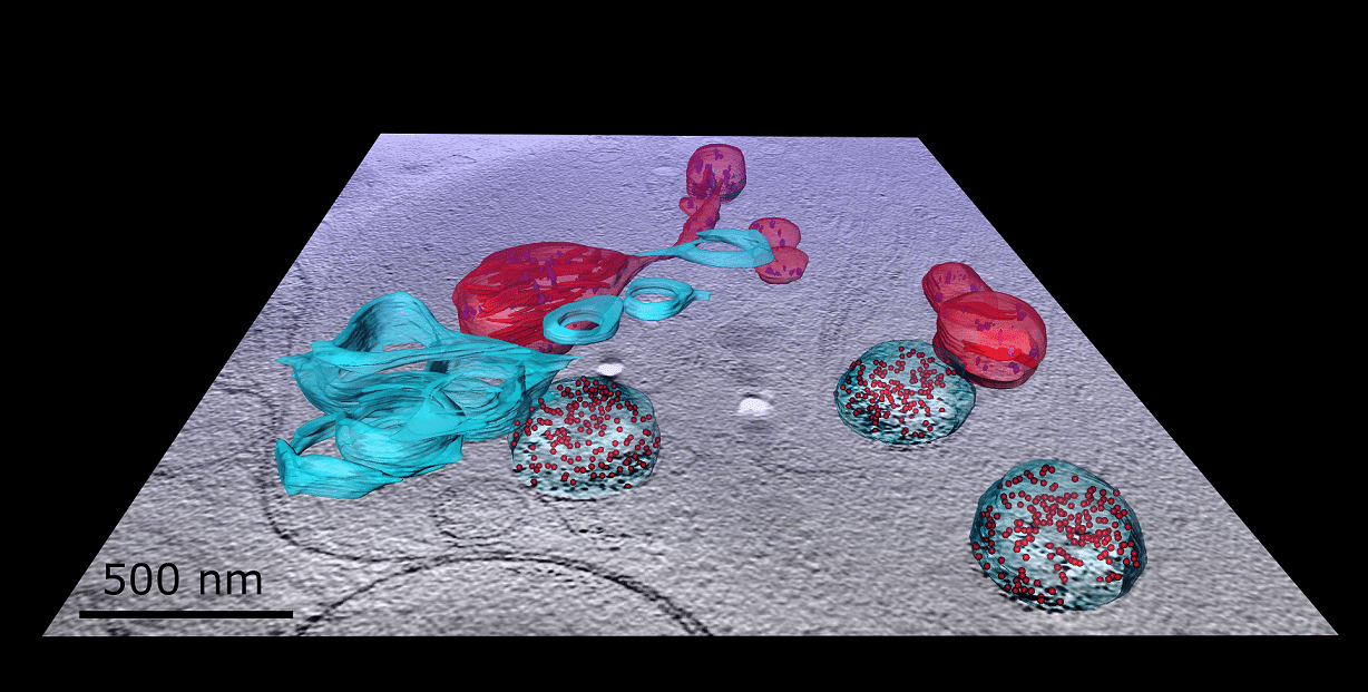
Segmentation of cryo-STEM tomogram coming out of a z-slice from the tomogram. Image shows a portion of a WI fibroblast cell containing mitochondria (red), membrane formations (blue), and intake vesicles (blue spheres) filled with gold-labeled collagen VI (red dots).