OBJECTIVES: To assess the effects of continuous positive airway pressure (CPAP) treatment on brain structure and function in patients with obstructive sleep apnea (OSA).
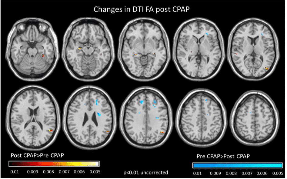
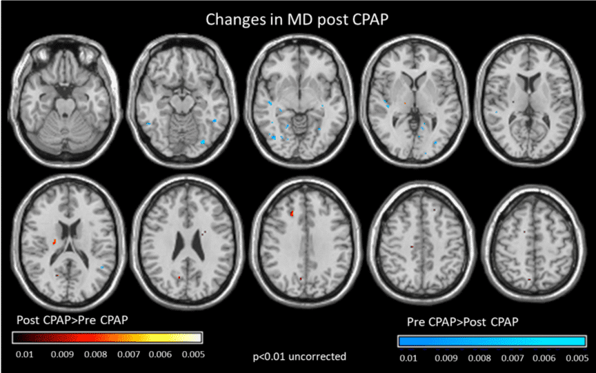
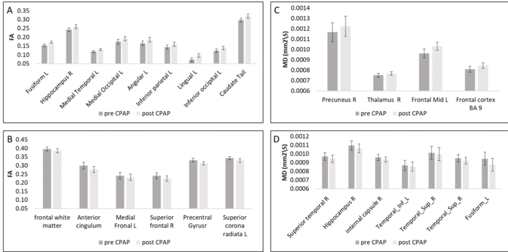
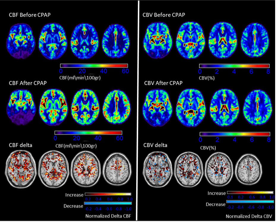
METHODS: A prospective study of seven OSA patients recruited from the sleep center at our institution. Patients were treated with six weeks of CPAP treatment. Pre-treatment and post-treatment MRI perfusion scans were obtained and compared to assess for treatment-induced changes. Microstructural changes were quantified using functional anistrophy (FA) and mean diffusivity (MD), and brain perfusion was quantified using cerebral blood flow (CBF) and cerebral blood volume (CBV).
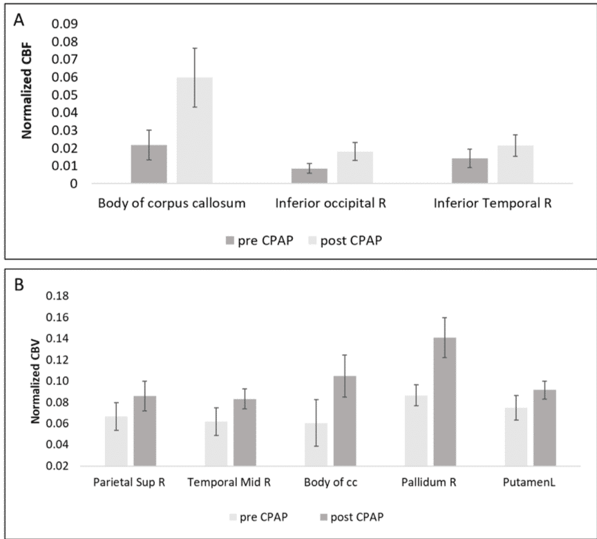
RESULTS: Of the seven patients included the in study, 6 (85.7%) were male and the mean age of the study population was 51 years (standard deviation = 13.14). Increased FA and decreased MD were found in the hippocampus, temporal lobes, fusiform gyrus, and occipital lobes. Decreased FA and increased MD were found in frontal regions for all patients (p <.05). Increased CBF and CBV were also observed following treatment (p <.05).
CONCLUSION: In addition to symptom resolution, CPAP treatment may allow for healing of OSA-induced brain damage as seen by restoration of brain structure and perfusion.