PUSPOSE: Salivary glands pathologies may be diagnosed using sialography - Cone-Beam Computerized Tomography (CBCT) imaging of the salivary glands following the introduction of iodine contrast solution into the salivary gland`s orifice. However, there is currently no definition of what constitutes a normal salivary-gland architecture. We hypothesize that the quantitative analysis of salivary gland model obtained from normal parotid Sialo-CBCT scans will help to characterize the normal glandular structure and improve the diagnosis of abnormal conditions.
METHODS: We have developed a method for segmentation and modeling of the parotid salivary ducts. The input is Sialo-CBCT scans considered normal by experts` opinion; the output is a segmentation of each of the salivary ducts and a model and analysis of the glandual ductal system. The algorithm is fully automatic and consists of two phases: segmentation and modeling. Segmentation includes Region of Interest (ROI) extraction, detection of tubular structures by Hessian filtering and geometric analysis, level-set segmentation, and identification of the tree-like salivary ducts structure. Modeling includes segmentation skeletonization to obtain a graph with nodes and edges, identification of the tree root, extraction of its branches and levels (orders) and duct model characteristics extraction.
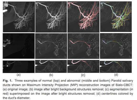
RESULTS: We defined a partial, semi-quantitative evaluation method for our algorithm based on Maximum Intensity Projection (MIP) images with the segmentation results superimposed on them. The evaluation is performed by assigning the first and second level branches grades between 0- and 4 (0 -- branch not detected, 4 -- branch fully detected), and counting how many of bifurcations were found in those branches. Based on this scoring, we compute the percent of the ducts branches and bifurcation were detected by our algorithm.
We evaluate our method on 13 Sialo-CBCT which were considered normal of different patients. It successfully identified 92% of the gland`s ducts; the rest were not identified mostly because of scan artifacts or because of their small diameters. For the successfully identified ducts, an average of 96% (std=14) of the primary ducts and 85% (std=22) of the second level branches, were correctly detected, based on visual inspection. The average detection rate for the second level branches was 66% (std=25) and 55% (std=32) for the bifurcations.
CONCLUSION: Automatic computer-based image analysis diagnosis of patient Sialo-CBCT scans may be a useful tool for the characterization of normal salivary gland architecture, which will further aid in diagnosis of glandular pathologies.
KEYWORDS: segmentation, salivary gland imaging, sialography, sialo-CBCT