
Significance of Long Phase Venography in CRT-D Implantation in a Patient with Persistent Left Superior Vena Cava
Aim
We would like to report a case of 75 years old man in whom LPVCS was overlooked due to inadequate interpretation of venography and emphasize the significance of long-phase venography depicting the full course of the subclavian vein.
Case Report
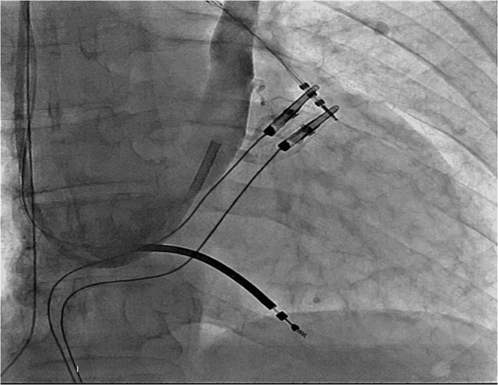
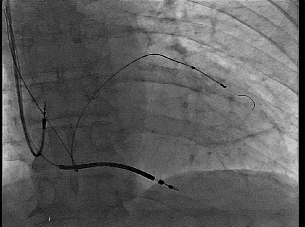
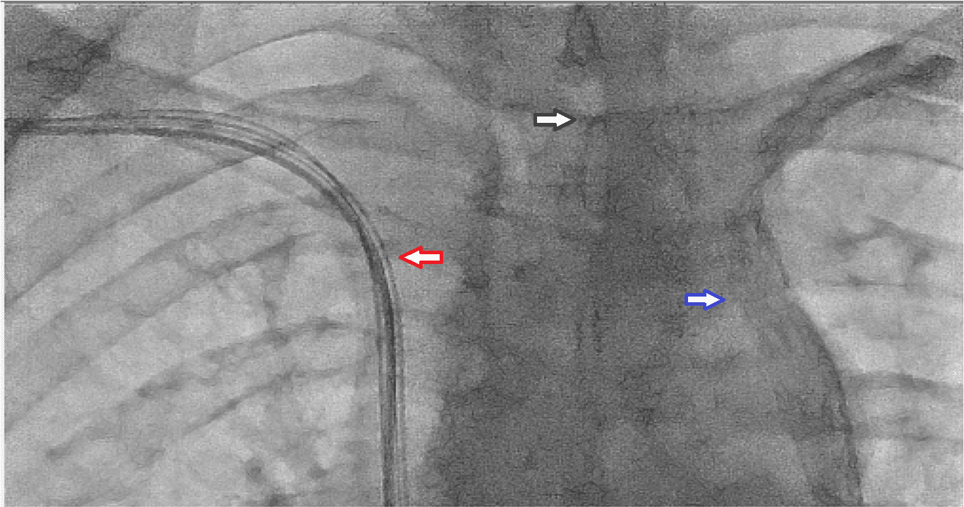
A 75 years old man with an ejection fraction of 35%, QRS duration 155 miliseconds and LBBB morphology on surface electrocardiogram was shedduled for CRT-D device implantation. Venography was performed as a part of our routine practice in order to explore the patency of the subclavian vein. After being sure about the patency of the vein, a pocket was created in the left pectoral region. The vein was canulated but the attempt to advance the guidewire has aborted. The tip of the guidewire fastened on a steep angle through the course of the vein. The venography was looked on again. Presence of a regressed innominate vein in a tapered aspect was noticed as well as a downward flow on the left side of the view . The patient had a PLVCS. Venography using the right antecubital vein showed the presence of a patent right SVC. A new pocket was created on the right pectoral region. Atrial, right ventricular and left ventricular leads were implanted using the right axillary vein puncture (Figure 1, 2). The procedure was quite straightforward and carried out succesfully (Figure 3).
Conclusion
Regression of the innominate vein in a tapered aspect caused us to overlook the presence of PLVCS. Venography from the right antecubital vein depicted the presence of right vena cava superior emptying in right atrium. Here the significant point to emphasize is that long phase venography is crucial in every case not to overlook PLVCS especially in the presence of a regressed innominate vein in a tapered aspect or a bridging innominate vein.
Powered by Eventact EMS