PURPOSE: Accurate estimation of abdominal aorta aneurysm is a well-known problem to radiologists mostly due to fusiform structure. Follow-up for large aneurysms is usually done with CTA and therefore includes exposure to radiation and contrast material. Small aneurysm follow-up is usually done with US and therefore less precise. CT-US fusion has evolved in recent years as a new promising tool to improve accuracy of invasive US-guided procedures. We hypothesized that using this technique for measuring abdominal aneurysm size will have better accuracy and reliability compared to conventional US.
MATERIAL & METHODS: The study included 11 patients with infra-renal aortic aneurysm and recent abdominal CTA. Each of the patient had a conventional US exam to estimate the aneurysm size and immediately after, a CT-US fusion exam to measure the aneurysm again, this time based on registration to the recent CTA (Fig. 1). Gold standard values for aneurysm size were extracted from the CTA using PORTAL-PHLIPS and marks of the maximal diameter were added to the DICOM files using MATLAB. The measurements of the fusion and conventional ultrasound were compared the CTA values.
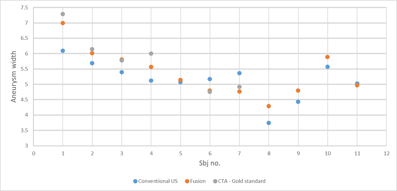
RESULTS: Despite the small sample, fusion measurements were more accurate and reliable than the standard examination (Fig. 2). Specifically it seems that good registration between the CTA and the US allow better estimation of fusiform aneurysms using US.
CONCLUSION: Our results suggest that CT-US fusion may prove to be a better study for follow-up of small abdominal aortic aneurysms. A larger cohort is needed to validate this result and to check whether the fusion technique can replace the CTA for follow-up of larger aneurysms, hence reduce exposure to radiation and contrast material.
Fig 1:
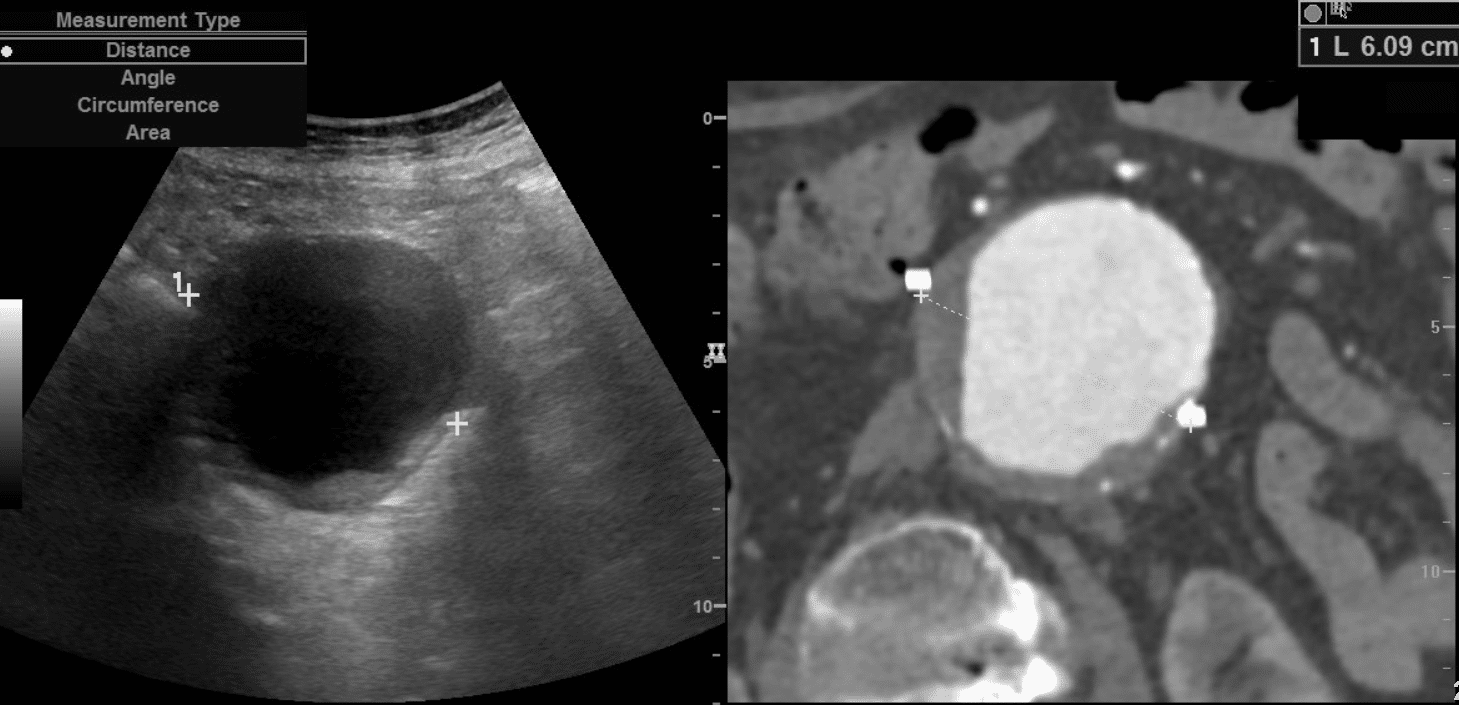
Fig 2 (not completed):