
Optogenetic Control of Spatial Heterogeneity in Human Induced Pluripotent Stem Cell Derived Cardiac Tissue
Aims:
The morphology and spatial distribution of the action potential (AP) in the heart are carefully controlled, and any deviation increases the risk for arrhythmia. Here, we aimed to use optogenetic technology to shape the AP morphology in human multicellular tissue models, to induce different spatial patterns of repolarization, and to evaluate their role in arrhythmia formation.
Methods and results:
293HEK cells were engineered to express the light-sensitive cation channel CoChR. The engineered cells were co-cultured with human induced pluripotent stem cells derived cardiomyocytes (hiPSC-CMs) to allow formation of gap junctions and electrical coupling. Optical mapping was used to monitor the electrical activity of the co-cultures. Optogenetic stimulations were patterned at high spatiotemporal resolution using a digital micromirror device.
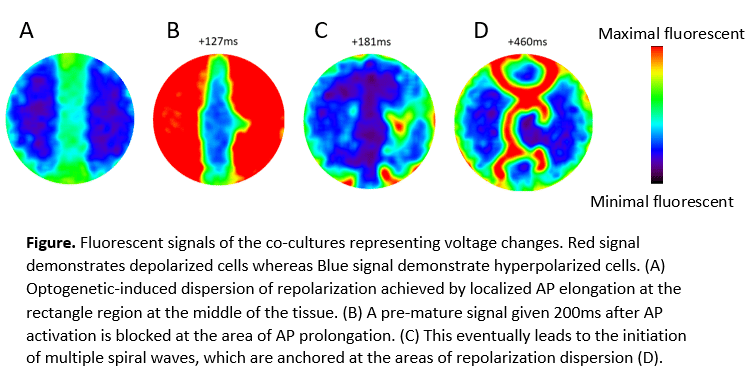
Light stimulation significantly prolonged AP duration (APD) in the cultures by 30%-50% (p=0.0044). Moreover, by spatially patterning the optical stimulation we produced APD gradients within the tissue (Figure A,B). APD prolongation of 50-100ms at selected illuminated areas increased the dispersion of repolarization and significantly increased the tissue vulnerability to develop arrhythmia. Hence, 83.33% of premature stimulation protocols induced arrhythmia when dispersion of repolarization was increased, relative to 25% at baseline (p=0.0123). Moreover, the time window for arrhythmia formation was increased: premature stimulation induced arrhythmia when delivered 200-250ms after AP stimulation when dispersion of repolarization was increased, compared to 200ms at baseline. The generated arrhythmias were reentrant in nature (rotors) and were anchored to the areas of repolarization dispersion (Figure D,C).
Conclusions:
Optogenetics interventions can be used to shape AP morphology in human multicellular model. Such interventions allowed to create and study the effects of localized areas of repolarization gradients. The increased variability in APD increased arrhythmogenicity by facilitating development of stable rotors. These results highlight the potential of combining optogenetic and hiPSC technologies in providing mechanistic understanding of arrhythmogenicity.
Powered by Eventact EMS