Beyond POCUS: Bedside Ultrasound in the Diagnosis of Soft Tissue Mass

Eric Scheier Uri Balla
Pediatric Emergency, Kaplan Medical Center, Israel

Point of care ultrasound (POCUS) has become an integral part of pediatric emergency care. ACEP states that “US [ultrasound] fulfills the concept of ‘staged imaging,’ where the use of US first can answer important clinical questions accurately without the expense, time, or side effects of advanced imaging or invasive procedures”. There exist a number of case reports describing the rare POCUS diagnosis of cystic structures and solid masses. However, the literature on the POCUS diagnosis of solid masses remains sparse. To date, no case series exists describing the evaluation of solid masses by POCUS in a single center.

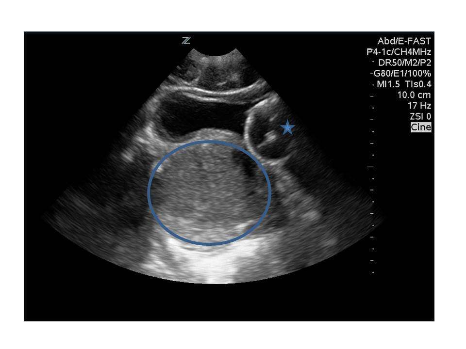
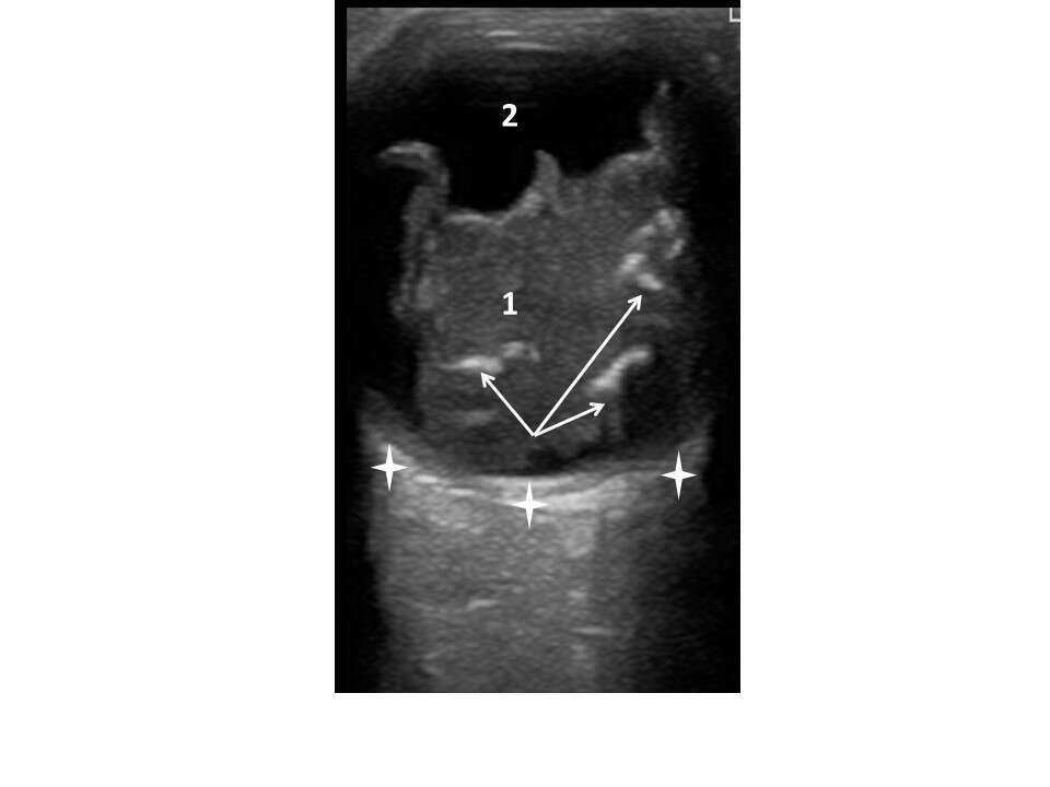
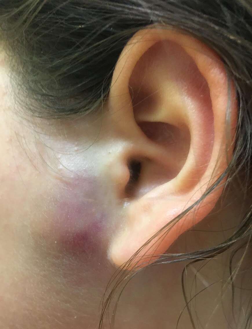
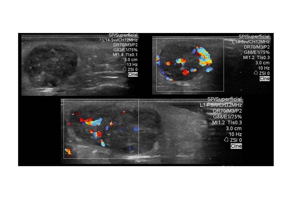
Here, we report six cases of a mass identified by the author, a novice sonographer in a pediatric emergency medicine fellowship, using POCUS, from January of 2017 to May of 2018. Identification of these cases early in presentation allowed earlier involvement of the relevant specialist, shorter time to diagnosis and treatment, and can be expected to increase parent satisfaction as well.

Transverse view of the rhabdomyosarcoma beneath the bladder. The mass is marked with a circle. The catheter balloon is marked with an star.Transverse view of the retinoblastoma. The mass is marked by the number 1, the vitreous is marked by the number 2, the calcifications within the mass are indicated by the arrows and the fundus is marked by stars. Note the point of attachment of the mass to the fundus on the left.Photo of preauricular mass.Top left, transverse view showing a heterogenous round subcutaneous mass,  suspicious for abscess. Top right: The same view with doppler shows a vascular mass inconsistent with abscess. Bottom: Coronal view of mass with Doppler.Phased array, right upper quadrant mass. The right upper quadrant mass is marked by the number 1. The kidney is incompletely viewed and marked by the number 2. An arrow marks the diaphragm.US views of right liver and kidney showing the communication of the abdominal mass with the right kidney. Liver is marked by an asterisk, kidney by a triangle, and the mass by a circle.


Powered by Eventact EMS