The immobile arm after a fall: a POCUS screening protocol to identify fracture in children with upper extremity injury.

Eric Scheier Uri Balla
Pediatric Emergency, Kaplan Medical Center, Israel

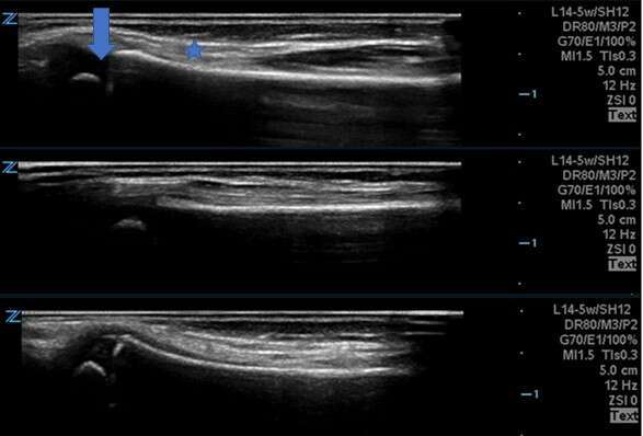
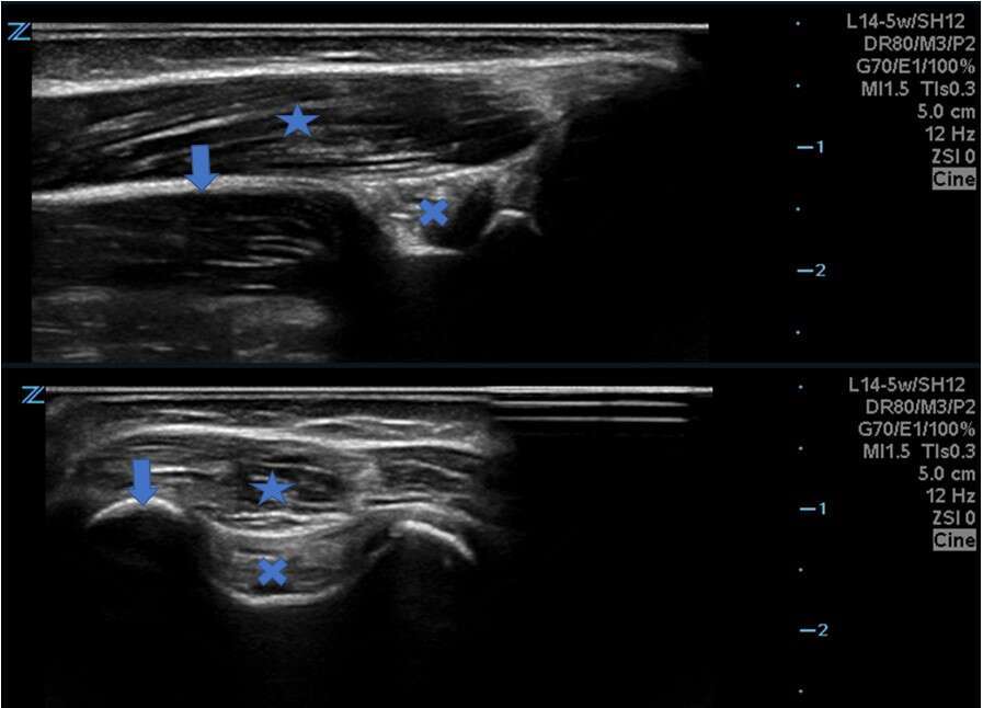
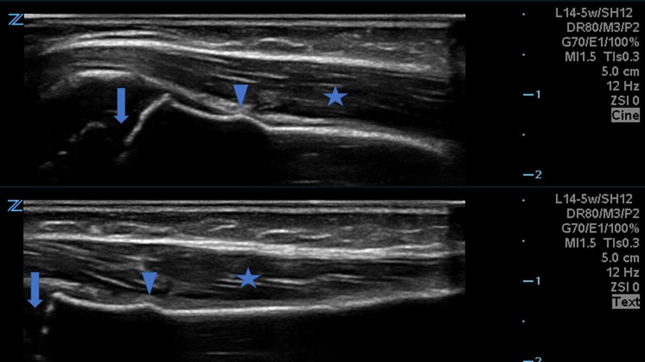
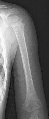
Pediatric emergency department physicians are often unable to discern a specific point of tenderness when examining children who present after minor trauma. Traditionally, the injured child without focal findings on history or examination is sent for radiography of the injured limb. Here, we report a case of a three year old boy who, while running, fell on an outstretched arm. On point of care ultrasound (POCUS) distal forearm, supracondylar area and proximal humerus were interrogated, and a torus fracture of the proximal humerus was found. In injured children without localizing findings on examination, a step-by-step POCUS screening protocol can quickly and painlessly identify common pediatric fractures.

Top: Dorsal radius. Middle: coronal radius. Lower: palmar radius. The probe indicator is at the distal end of the bone. On the image at the top, an arrow indicates the distal physis, and a star indicates muscle overlying the white hyperechoic border of bone.

Top: sagittal view of posterior distal humerus. Bottom: transverse view of posterior humerus. Probe indicator is proximal on the top image and lateral on the bottom image. An arrow indicates bone: distal humerus in the top image and lateral condyle in the bottom image. An X indicates a normal fat pad in the olecranon fossa. A star indicates triceps.

Top: Coronal view of proximal humerus. Bottom: Posterior sagittal view of the proximal humerus. Probe indicator is proximal. an arrow indicates the proximal physis, a triangle points to the fracture site and a star sits in the triceps muscle.AP view, torus fracture of the proximal humerus.


Powered by Eventact EMS