
Non-Invasive Thermal Imaging Identifies Left Ventricular Remodeling in Mice
2Neufeld Cardiac Research Institute, Tel Aviv University
3Tamman Cardiovascular Research Institute, Sheba Medical Center
4Internal Medicine Wing, Sheba Medical Center
5School of Medical Engineering, Afeka Tel Aviv Academic College of Engineering
6School of Electrical Engineering, Afeka Tel Aviv Academic College of Engineering
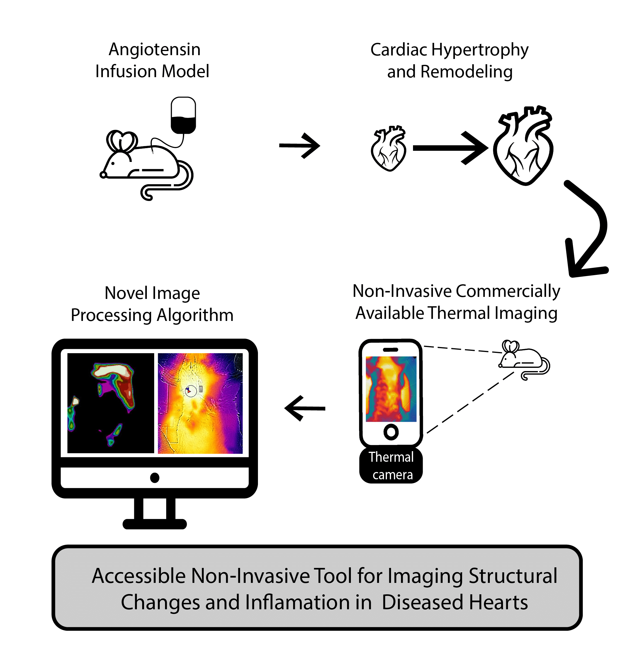
Background: Thermal infrared imaging is a non-invasive alternative to monitor physiology and disease. The use of this technique to image internal organs, such as the heart, has not yet been investigated. We aimed to determine the ability of a novel algorithm for thermal image-processing to detect structural and functional changes in a mouse model of cardiac remodeling.
Methods: We randomized 12 male mice (weight 20-25 gr) to treatment with either angiotensin-II (2 mg/kg/day, n=6) or saline (n=6) pump-infusion for 28 days. We measured blood pressure weekly, together with serial trans-thoracic echocardiography studies and histopathological evaluation of the hearts. We captured thermal images with the commercially available FLIR One camera, and processed images by our novel algorithm.
Results: Angiotensin infusion increased blood pressure together with cardiac hypertrophy and fibrosis. Thermal imaging identified an increase in the fraction of the skin heated by the heart in angiotensin-treated mice, at day 28 of the experiment. Thermal image findings were correlated to left ventricular mass and volume by echocardiography (r= 0.6, p=0.07 and r=0.8, p< 0.01). By thermal imaging, all angiotensin-treated hearts displayed a unique triangle-like shape of heat distribution. This finding was absent in controls, indicating remodeling in the hypertensive heart.
Conclusion: Our preliminary findings suggest, for the first time, that a thermal camera with a new image-processing algorithm, identifies cardiac structural changes in mice. Our findings propose a new, simple, and non-invasive tool to diagnose and monitor adverse cardiac remodeling.
Key Words:
Angiotensin; Cardiac hypertrophy; Image processing; Mice; Thermal imaging
Visual Abstract:
Powered by Eventact EMS