
BrainWatch: Brain Damage Alert by Continuous Pupillary Monitoring Through Closed Eyelids
2The Hebrew University of Jerusalem, Israel
3Hadassah Ein-Kerem University Hospital, Israel
4The Hebrew University of Jerusalem, Israel
5Hadassah Ein-Kerem University Hospital, Israel
The pupillary light reflex and pupil size are important clinical parameters and prognostic indicators in the evaluation of critically injured or comatose patients. The Brain Trauma Foundation and the American Association of Neurological Surgeons recommends that pupillary light reflex for each eye should be used as a prognostic parameter and that the duration of pupillary dilation and fixation should be documented.
Currently, most pupil measurements are conducted the nursing staff by manually opening the patient’s eyelids and using a ruler and a flashlight to measure pupil size and light reaction, respectively. This measurement is infrequent, inaccurate and increases staff workload. Here we describe a continuous, accurate, standardized, automatic and hands-free wearable device capable of measuring pupil size and light response through closed eyelids. The device is based on near-IR illumination and imaging to measure pupil size, and visible light illumination to excite measurable pupil contraction. The device will enable real-time alert on changes in the patient’s status. First results on 40 healthy volunteers show 97% success rate in measuring pupil size and light response (figure 1).
In comatose patience, examination of the pupils is crucial for early detection of various medical events. Currently, this examination is performed manually and infrequently. Here, for the first time, we present an automated pupil measurement through closed eyelids, which will potentially improve the standard of care.
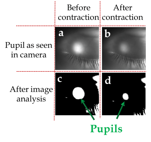
Figure 1: Pupils can be identified through closed eyelids, (a) before white light exposure, (b) after white light exposure. (c) and (d) image analysis for both acquisitions.
Powered by Eventact EMS