
Detection of Apoptosis with 99mTc-rhAnnexin V-128 Imaging in Asymptomatic Patients with Carotid Atherosclerosis Plaque
Background: Apoptosis plays an important role in the late progression of atherosclerotic plaque and may be useful for identifying advanced and complicated lesions.
Methods: Proof of concept, Phase II study.
Patient recruitment from the carotid ultrasound laboratory.
Carotid artery (CA) ultrasound within 8 weeks of the annexin scan,
Significant CA disease (≥ 50% stenosis in one or both CAs.
Exclusion: Previous CA intervention, stroke or TIA.
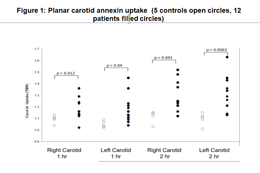
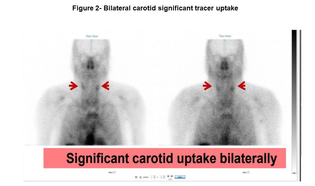
planar and SPECT imaging (GE Hawkeye SPECT/CT) at 1 and 2 hours after intravenous injection of ~ 350 MBq of 99mTc-rhAnnexin V-128. Images were reviewed by 3 expert reviewers for identification of focal uptake in the CAs and in the aorta.Quantitative analysis of the planar images was carried out by placing ROI in the CA and subclavian region representing venous and arterial activity (target to background (TBR)).
Results: 582 patients screened, 21 recruited and 17 completed the protocol (5 normal controls and 12 patients. No adverse events. One patient of 12 (8%) had significant focal uptake in both CAs at 1 and 2 hours. No uptake seen in the CAs of the other 11 patients or in the 5 controls (0/5, 0%). Carotid uptake of annexin was significantly greater in the patients with CA disease compared to controls for both CAs at 1 and 2 hours (RtCA at 1 hour: 1.18 ± 0.09 vs 1.09 ± 0.03, p=0.012; LtCA at 1 hour: 1.18 ± 0.12 vs 1.05 ± 0.03, p=0.04; RtCA at 2 hours: 1.29 ± 0.12 vs 1.12 ± 0.05, p=0.001; LtCA at 2 hours: 1.32 ± 1.14 vs 1.1 ± 0.05, p=0.0002).
Conclusions: 99mTc-rhAnnexin V-128 imaging of apoptosis is feasible with excellent image quality. Significant focal uptake was identified in 1/12 patients and 0/5 of the controls. Quantitative analysis of the planar images showed significantly greater uptake in patients compared to controls.

Powered by Eventact EMS