
The Synergistic Effect of Low-Frequency Ultrasound Combined with Spironolactone for Reduction of Fibrosis after Myocardial Infarction in Rats
2Eliachar Research Laboratory, Galilee Medical Center
3Department of Internal Medicine F, Galilee Medical Center
4The Azrieli Faculty of Medicine, Bar-Ilan University
Background: Myocardial infarction (MI) is a major cause of myocardial collagen deposition leading to left ventricular remodeling and heart failure. The renin-angiotensin-aldosterone system (RAAS) plays a major role in ventricular remodeling. Post MI, spironolactone attenuates cardiac remodeling and subsequent fibrosis. We hypothesized that non-invasive transcutaneous therapeutic ultrasound (TUS) at a frequency of 1 MHz and intensity of 0.5 W/cm2 with the addition of an aldosterone inhibitor may affect collagen deposition early after MI.
Methods: After echocardiographic evaluation rats underwent MI induction procedure. Subsequently rats were treated either by daily spironolactone, TUS (3 times a week) or their combination. Control rats were not treated. We re-evaluated cardiac function two weeks after treatment initiation and at the end of the experiments (4 weeks). We performed histological and biochemical evaluation of fibrosis after sacrifice.
Results: The basal ejection fraction (EF, 70±2%) deteriorated following MI induction to 36±3% (Pvs. baseline). TUS treatment alone did not significantly improve EF 4 weeks after MI (45±3%). Spironolactone alone increased the EF to 50±2%, (Pvs. control). A synergistic effect was observed in the dual treatment as EF increased to 54±3% (Pvs. control). On histology, we found a significant reduction in collagen deposition (Pvs. control) following spironolactone or the dual treatment (Pvs. control).
Conclusions: This study demonstrates a positive effect of spironolactone on myocardial collagen content post MI, as well as a synergistic effect of spironolactone with transcutaneous TUS.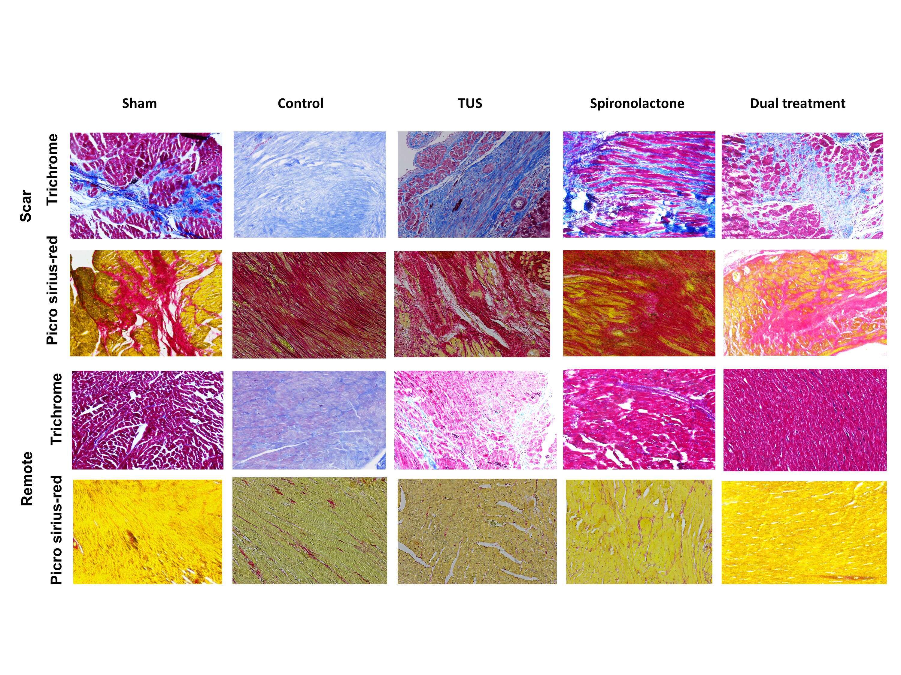
Figure 1: Representative histological specimens of cardiac muscles stained with Tri-chrome Masson`s (first and third rows), and Picro-Sirius red stain (second and fourth rows). Specimens in the first 2 rows were taken from scar areas, and the lower 2 rows were taken from remote areas. Each column represents the designated treatment. (X10 magnification).
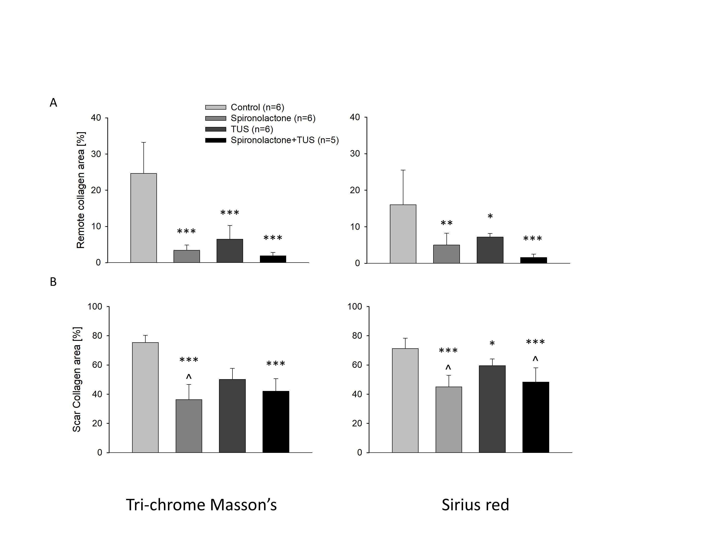
Figure 2: Quantification of collagen volume fraction in specimens taken from (A) Remote areas and (B) Scar areas. Left panel quantification from Tri-chrome Masson`s stained specimens. Right panel quantification from Picro-sirius red stained specimens. * Pvs. control, ** Pvs. control, *** Pvs. control.
Powered by Eventact EMS