
In-vitro Assessment of the Thrombolytic Efficacy of Therapeutic Ultrasound
2The Cardiology department, Galilee Medical Center
3The Azrieli Faculty of Medicine, Bar-Ilan University
Background: Ultrasound is mainly used as a diagnostic tool. Several studies demonstrated that therapeutic ultrasound (TUS) can enhance thrombolysis, but the optimal mechanical parameters to achieve this biological effect are still unknown.
Methods: We assembled 46 blood clots in a closed in-vitro circulatory model. Clots were randomly divided into 7 groups, control group and six TUS groups of three frequencies (0.3, 0.5, 0.7 MHz) and six intensities (0.75, 1.5, 3, 237.7, 475, 950 W/cm2). Treatment was composed of 12 repetitions, 5 min US application and 3 min pause, lasting 93 minutes in total. Clots’ weight and flow rate were measured before and after the treatment.
Results: Mean initial clot weight (0.318±0.129 g) and flow (0.53±0.31 ml/min) were comparable among the experimental groups. We found a final clot weights reduction (0.15±0.05, 0.16±0.06, 0.09±0.07, 0.21±0.09, 0.17±0.09, 0.17±0.07 and 0.18±0.02 g in groups 1 through 6, respectively) and a flow increase (30.61±19.76, 52.1±25.44, 28.78±8.15, 43.93±20.03, 40.86±18.25 and 45.10±22.20 ml/min in groups 1-6, respectively) in all TUS groups. Clot weight change (%) and flow increase reveals that the TUS profile f =0.5 MHz I=1.5 W/cm2 was most efficacious. In the control group, clot weight change was +6.3% of baseline and flow increase of 4.4% of baseline, whereas -75.4% of baseline and 209.3% of baseline in the f=0.5 MHz I=1.5 W/cm2 profile were noted, respectively.
Conclusions: Our study proved that TUS augments thrombolysis in-vitro. TUS at low frequency (0.5 MHz) is most effective, whereas changing the intensity of TUS has only a minor effect on the clot lysis magnitude.
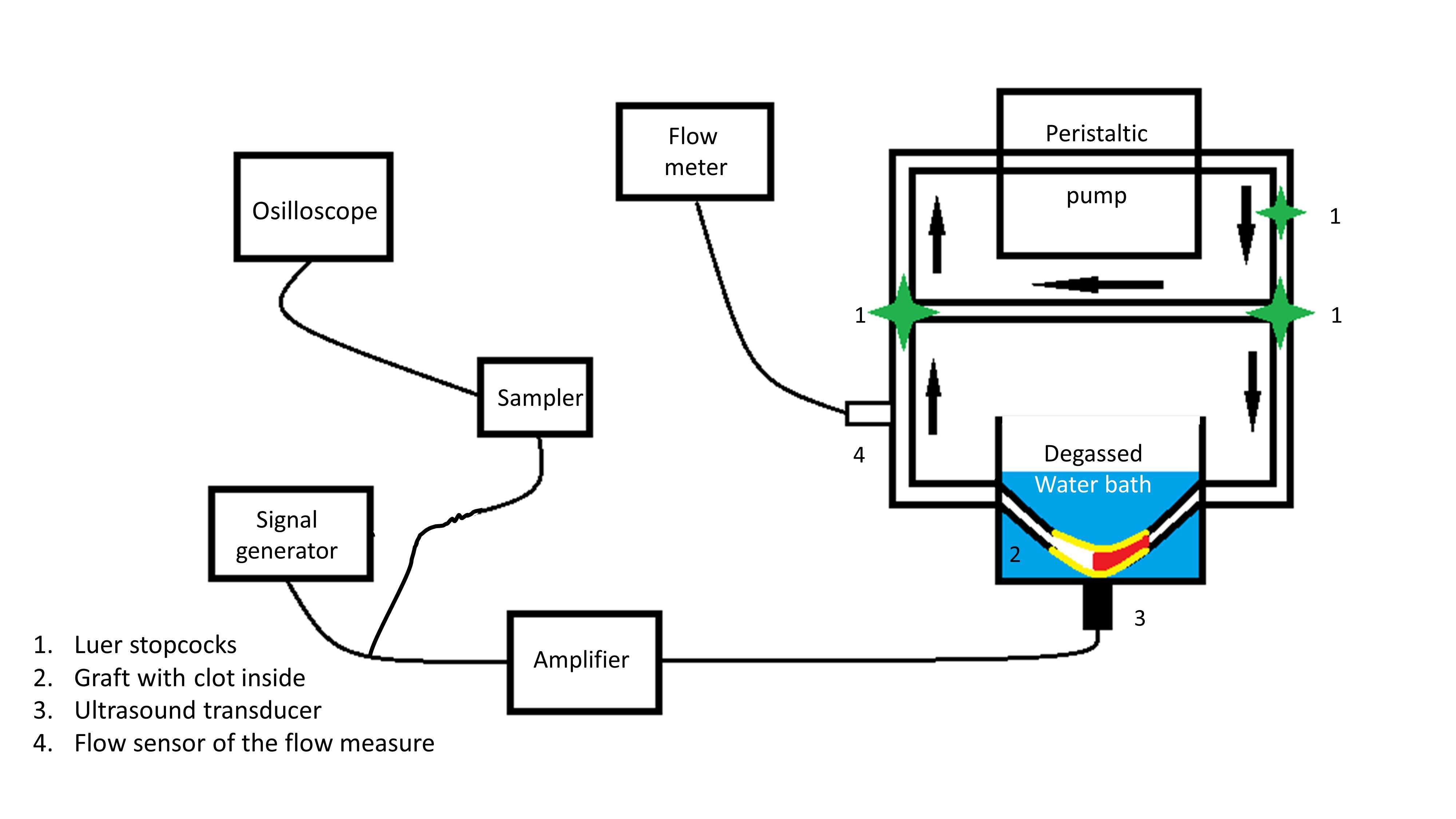
Figure 1: The experiment set-up
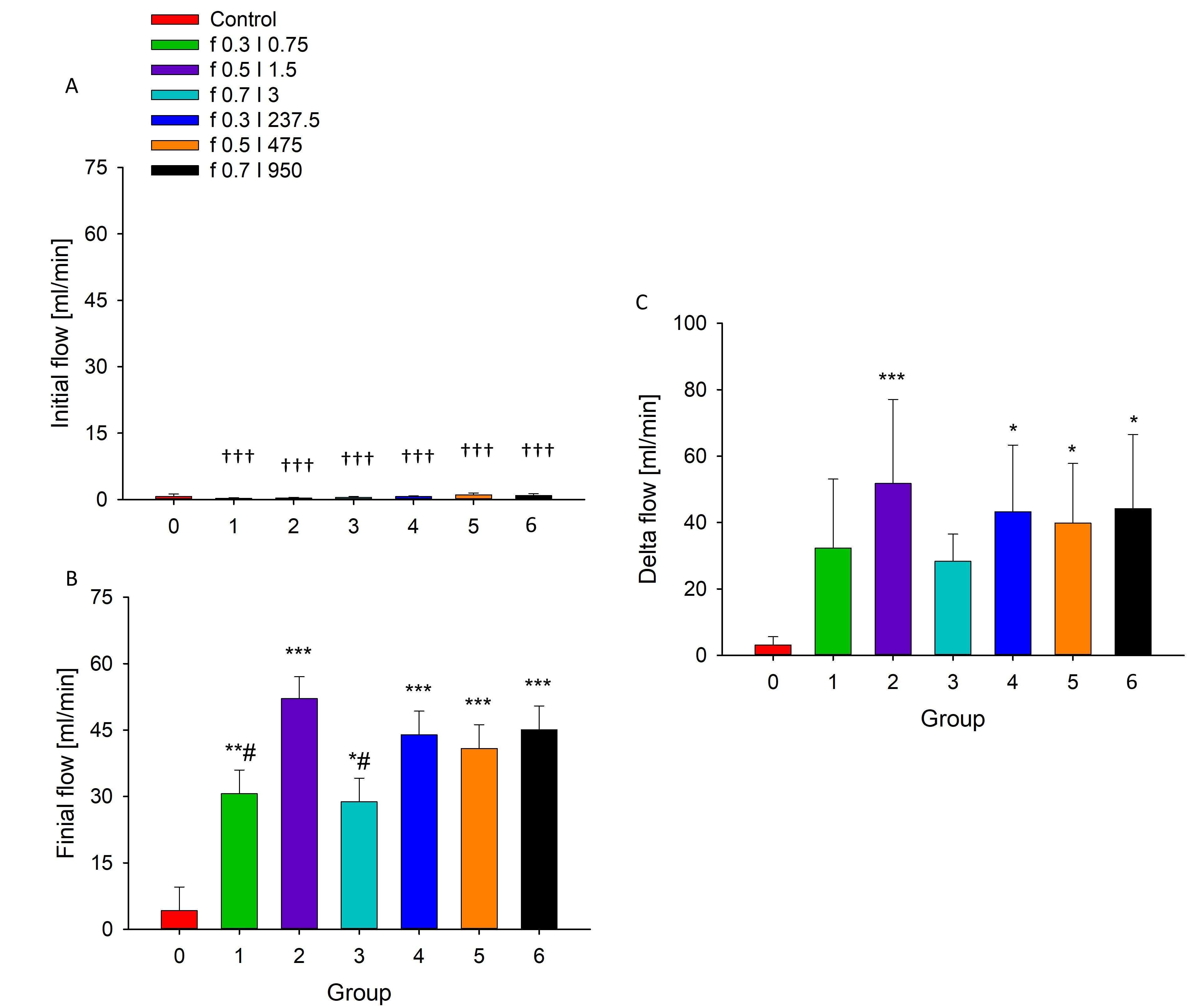
Figure 2: Saline flow in the set-up. A. initianl flow in the set-up, B. after treatment, C. The flow changes. ††† P<0.001 vs. post-treatment (A vs. B, in the same treatment group). * Pvs. control group, **Pvs. control group, ***Pvs. control group. # P

Powered by Eventact EMS