
Left Ventricular Circumferential Rotation is Associated with Functional Mitral Regurgitation
2Radiology, Shaare Zedek Medical Center
3Cardiology, Soroka University Medical Center
4Cardiology, Padeh Poria Medical Center
Background: Patients with left ventricular (LV) dysfunction may suffer from Functional Mitral Regurgitation (FMR). Impaired circumferential LV movement may influence FMR. We hypothesized that FMR is associated with LV systolic twist and torsion, evaluated utilizing novel cardiac Magnetic Resonance Imaging (MRI) processing techniques.
Methods: Normal subjects (n=49, mean age: 43y, 37% females, mean EF: 63%); patients with low EF (EF<50%) without FMR (n=32, mean age: 57y, 19% females, mean EF: 38%), and with FMR (regurgitant volume (RVol)>10 mL; n=37, mean age: 55y, 32% females, mean EF: 34%) were compared. All subjects underwent cardiac MRI for clinical indications. 2D tissue-tracking analyses were conducted using Circle CVI42 version 5.9.4 software. Systolic twist was calculated as the difference between apical to basal circumferential displacement. Systolic torsion was calculated as twist divided by the distance between apical and basal slices. RVol was calculated as the difference between stroke volume and the forward flow in the ascending aorta. Specific manually derived LV global and local measurements as well as mitral deformation parameters were evaluated. Groups were compared using ANOVA and correlations by Spearman’s test.
Results: Patients with FMR had significantly lower peak systolic twist (mean±SD 3.14±2°) and peak systolic torsion (0.55±0.38°/cm) compared to the patients with low EF (twist:5.7±2.25°, p<0.001; torsion:0.88±0.51°/cm, p=0.01) and to normal subjects (twist:5.57±1.9°, p<0.001; torsion:1.02±0.47°/cm, p<0.001). Among the patients with FMR RVol did not correlate with LV volume, function or annular diameter and weakly correlated with systolic separation between papillary muscles (r=0.44, p=0.03) and with lateral displacement of posterior papillary muscle (r=-0.52, p<0.01). RVol significantly correlated with peak systolic twist (r=-0.74, p<0.001; Figure 1) and peak systolic torsion (r=-0.55, p<0.001).
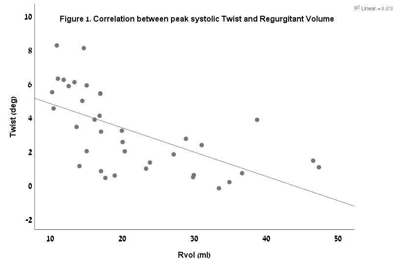
Conclusion: FMR severity is significantly associated with left ventricular twist, suggesting an additional pathophysiological mechanism.

Powered by Eventact EMS