
Hypertrophyc Pyloric Stenosis: A Radiographic Finding
2Neonatology, Hospital Universitario La Paz, Spain
Introduction: Hypertrophic pyloric stenosis (HPS) is one of the most frequent causes of non-bilious vomiting in infants. The diagnose is usually carried out by ultrasonography and only a few cases diagnosed by simple radiography are reported in the medical literature.We present a case that was diagnosed incidentally by the “caterpillar sign”, a characteristic radiographic sign of HPS.
Clinical Case: A 2-month-old male patient was operated on for a cervical teratoma. Subsequently, he presented weakness of the oropharyngeal musculature and difficulties in swallowing as well as severe gastroesophageal reflux. He was under omeprazole, domperidone and continuous debit nutrition through a transpyloric tube. Despite the treatment, the patient was presenting frequent regurgitation and vomiting and a chest-abdomen x-ray was perfomed to check the correct position of the probe. The radiography showed a distended stomach with increased peristaltic activity and he was diagnosed of HPS (Figure 1). The diagnosis was confirmed by abdominal ultrasound (figure 2) and a submucosal pyloromyotomy was perfomed with successful evolution.
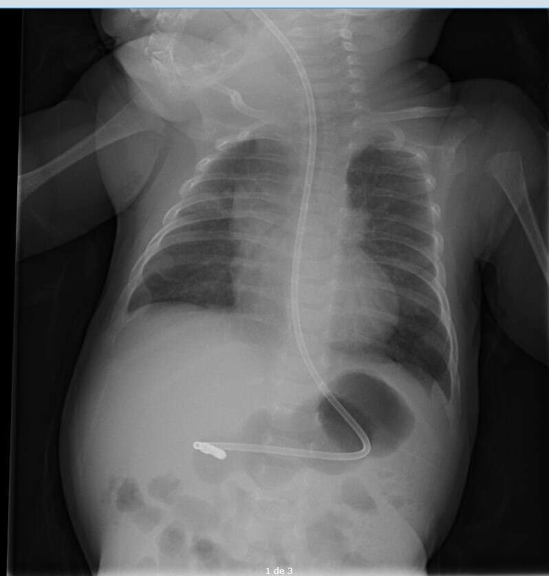
Figure 1. Chest-abdomen x-ray with notable peristaltic waves in the stomach due to HPS.
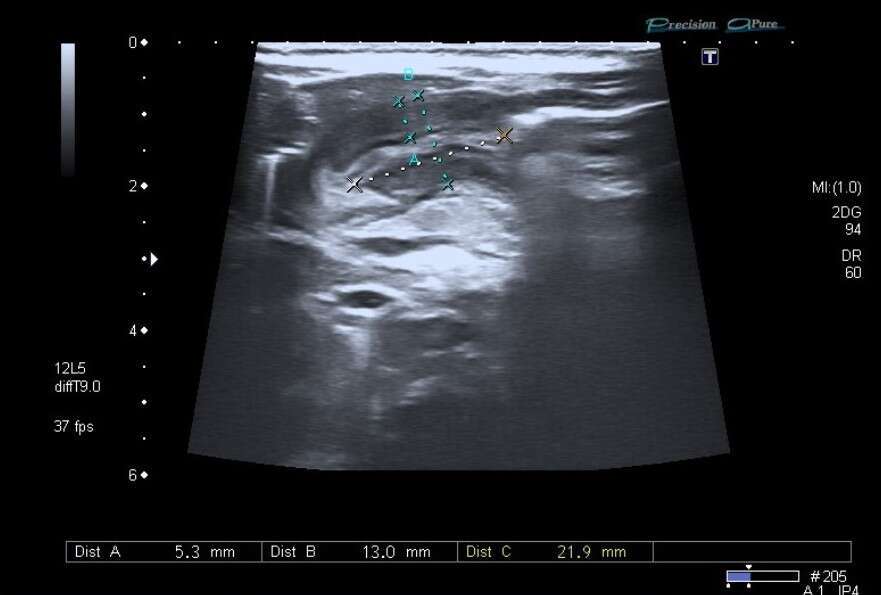
Figure 2. Abdominal ultrasound: Pyloric channel with diagnostic criteria for HPS.
Discussion: The gold standard imaging test for the diagnosis of HPS is abdominal ultrasound. If it does not provide a conclusive result, barium studies or upper gastrointestinal endoscopy are other diagnostic options.
Our patient was diagnosed incidentally by simple radiography of the abdomen by the "caterpillar sign". This sign is the result of vigorous peristaltic waves against an abrupt stop at the hypertrophic pylorus.
Conclusion: This clinical case reminds us the importance of the systematic analysis in medical imaging tests, particularly in simple radiography, as despite it being a basic diagnostic tool, it is also a great diagnostic source of information.
Powered by Eventact EMS