
Just another Case of Pneumonia?
Background: Pneumonia is a common infection but it can be the first presentation of an underlying disease, such as tumors.
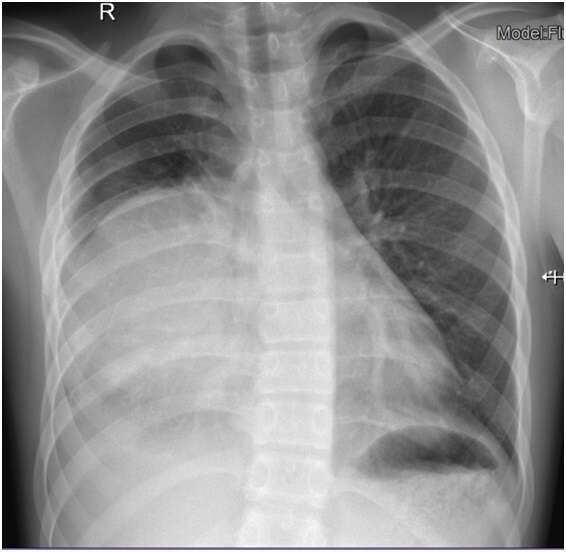
Case Summary Presentation: An 11 year-old boy, with atopic dermatitis, allergic rhinitis and asthma, presents to the emergency department with a 2 day history of thoracic pain accompanied with shortness of breath. Physical exam reveals fever and normal oxyhemoglobin saturation (95%–100%) on room air. On auscultation he has absent sounds in the right hemithorax but normal breath sounds on the left. A chest radiograph reveals a round area of consolidative opacity in the right hemithorax and a pleural effusion, confirmed by chest ultrasonography, with 14 mm. Blood tests show a leucocyte count of 16.600/mL with 78.3% of neutrophils, and an elevated C-reactive protein level of 17.59 mg/dL. He is admitted to the hospital and starts intravenous ampicillin. At day 2 after admission, the chest ultrasonography reveals a pleural effusion of 21.6 mm at the right hemithorax and images compatible with an empyema. He is submitted to a Video Assisted Thoracic Surgery (VATS) and drains about 100 mL of non-pure pleural fluid. The antibiotic therapy is changed to intravenous ceftriaxone. All cultural exams were negative. After 8 days, clinically improved, he is discharged and continues ceftriaxone until completing 14 days. One month later he repeats the chest radiograph which continues to reveal the same round area of opacity. The CT scan and the MRI show a round, encapsulated and cystic mass located in the anterior mediastinum compatible with a teratoma. He was submitted to excision of the tumor and pathologic evaluation revealed a mature teratoma. Subsequently, he is clinically improved and asymptomatic.
Conclusion: This case demonstrates that complicated pneumonia should always be followed up until complete resolution of symptoms and radiologic findings, and that a round consolidation should always be suspicious.
Powered by Eventact EMS