
Role of TGF-ß1/Smad Signaling Pathway in Vascular Injury of Retinopathy of Prematurity
2Neonatal Intensive Care Unit, Nanjing Children's Hospital, China
Objectives: To explore the changes of TGF-ß1/Smad signaling pathway in Phase 1 of retinopathy of prematurity and provide new targets for early intervention of ROP.
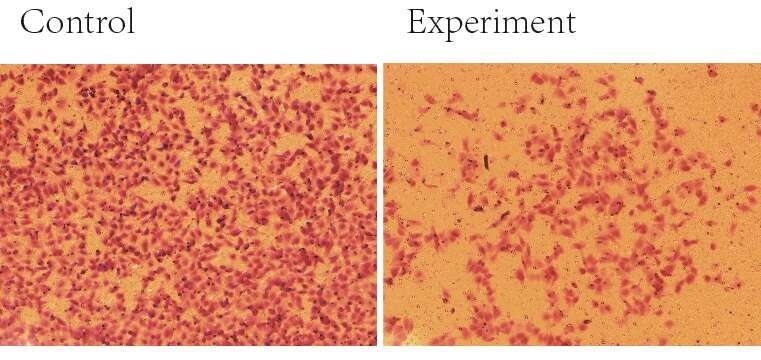
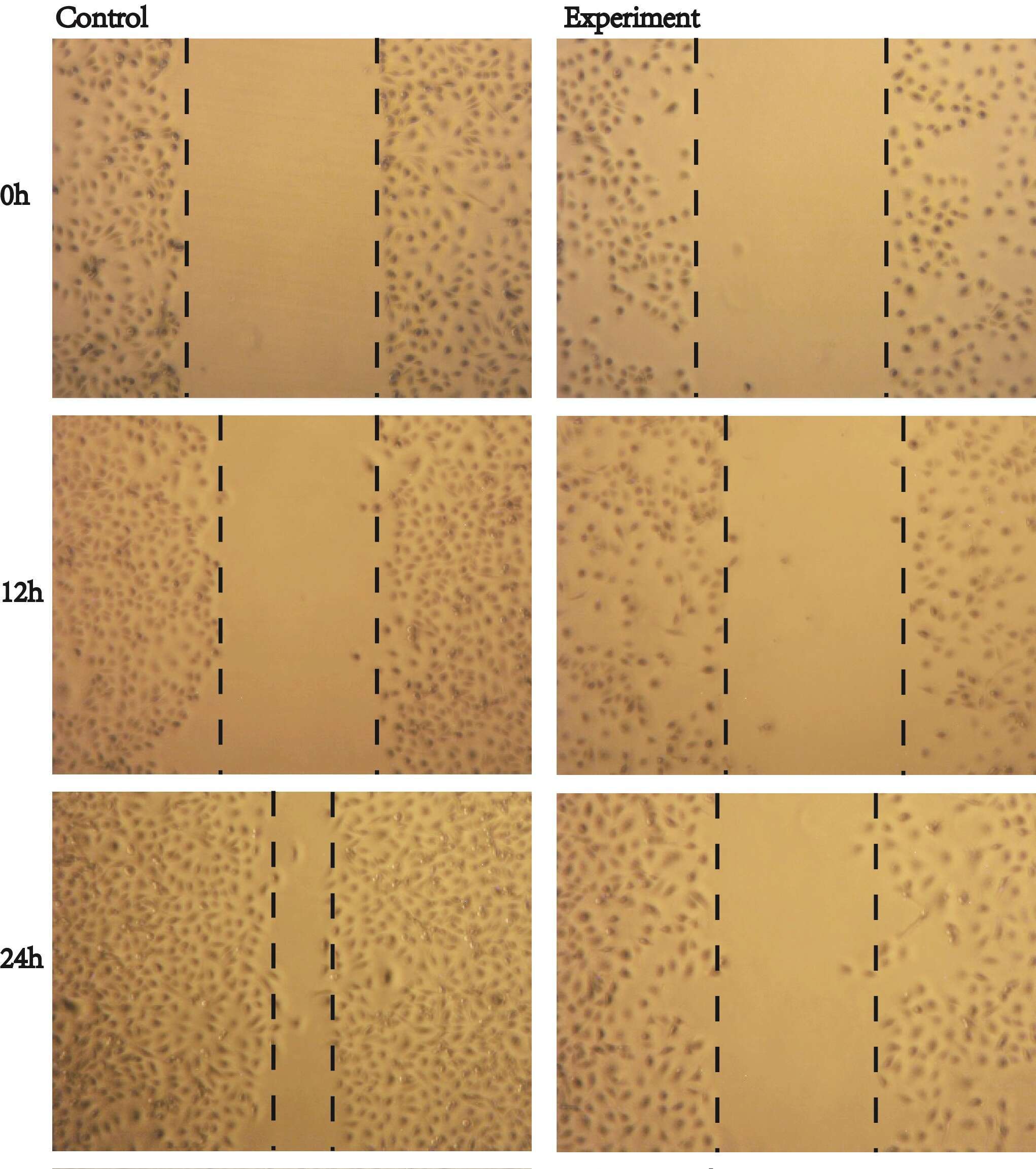
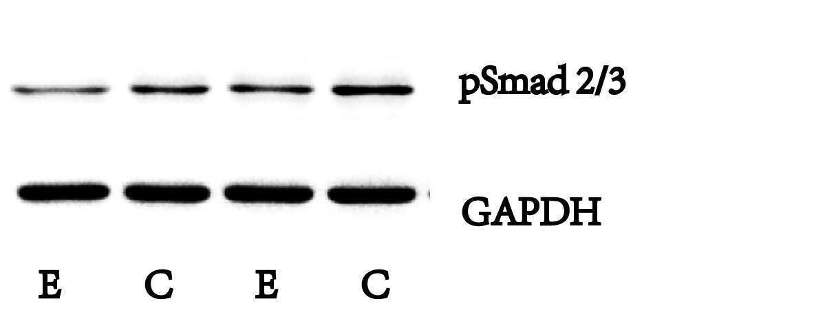
Methods: Human umbilical vein endothelial cells were cultured in vitro, and the experiment group was treated with hyperoxia-induced complete medium and hypoxia-induced complete medium intermittently. Wound healing kinetic and transwell migration assay were used to test the proliferation and migration abilities of HUVEC. RT-qPCR was used to detect the mRNA relative expression of TGF-ß1, and Western blotting was used to detect the expression of pSmad2/3 in cells of two groups.
Results:
1. After hyperoxia/hypoxia stimulation of HUVEC, the cell’s morphology changed, and the ability of proliferation and migration decreased.
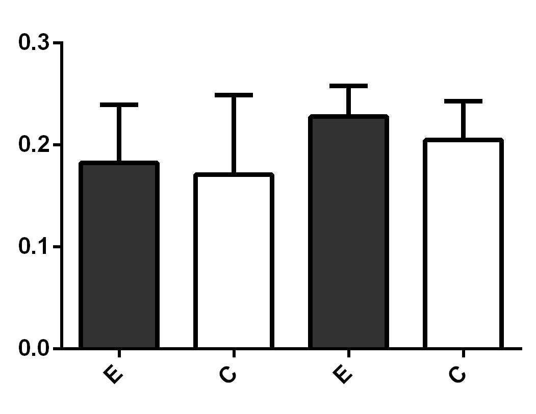
2. In RT-qPCR, the relative expression of mRNA in TGF-ß1 after stimulation was higher than that in the control group.
3. In Western blotting, after stimulation, the expression of pSmad2/3 in experiment group was lower than that in control group, and it was statistically significant.
Conclusions: In the hyperoxia/hypoxia HUVEC model, the expression levels of TGF-ß1 increased in experiment group. Moreover, the expression of pSmad2/3 was significantly reduced after the stimulation, along with the decrease of cell proliferation and migration ability, which indicated that the decreased of TGF-ß1/Smad2/3 signaling pathway would lead to the decrease of angiogenesis. The pathway may participate in the inhibition of angiogenesis in Phase 1 of ROP.
Powered by Eventact EMS