
Precocious Puberty Caused by Ovarian Dysgerminoma with Gonadoblastoma in a 46,XX Girl: A Case Report
2Endocrinology, Obstetrics and Gynaecology, Children's Clinical University Hospital, Latvia
Introduction: Gonadoblastoma is a rare tumour consisting of germ cells resembling granulosa and Sertoli cells. Sometimes these tumours contain stromal elements indistinguishable from lutein and Leydig cells. Almost all patients have 46,XY karyotype or various forms of mosaicism. Dysgerminomas are malignant, undifferentiated germ cell tumours that constitute 1% of primary ovarian neoplasms. Approximately 5% of dysgerminomas arise in abnormal gonads(from a gonadoblastoma).
Methods: The clinical case was analysed using patient history and clinical examination, laboratory, genetic, radiological and histological investigations, treatment methods and follow-up procedures.
Results: 6 years 4 months old female presented with early pubertal development, tall stature and a large abdominal mass. Height 135cm(+3.2 SDS) weight 30.45kg(+2.0 SDS). Tanner puberty stage III: Px4, Ax1, Ma3, Me1. Signs of virilization were present.
Laboratory data: significantly elevated estradiol, androstenedione, testosterone, alfa-fetoprotein, beta-hCG and AMH. LH and FSH – prepubertal level.
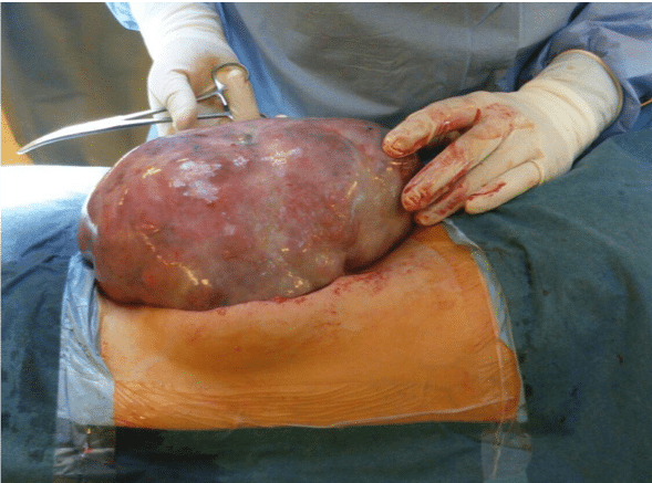
Bone age(RUS score-598 points) corresponded to an age of 12.2. MRI showed large heterogeneous mass(CC 25.1cm, LL 18.9cm, AP 10.3cm) on left ovary. Left periaortic lymph nodes(LN) were increased.
An operation was done after ovarian tumour was confirmed by MRI. Tumour and two additional LN were completely resected, biopsies were taken. Histological report: left ovarian dysgerminoma with metastasis in periovarian LN. However, the tumour was hormonally active, therefore, Sertoli cell component could not be excluded. Consultation from Finnish pathologists suggested dysgerminoma in combination with gonadoblastoma. Karyotype analysis of blood was 46,XX. Skin biopsy excluded mosaicism.
Four courses of chemotherapy were done. Further follow-up was provided by a multidisciplinary team. Blood analysis was taken to monitor hormone levels in each hospital visit. Skeletal scintigraphy, chest CT, abdomen MRI was done after chemotherapy – no metastasis was found.
Conclusions: A delayed precocious puberty diagnosis has caused this child significant health issues: hormonal disbalance, prognostic small adult height, therapeutical side effects from chemotherapy, increased risk for metastasis and secondary tumour development. For an early diagnosis of possible malignity, every child needs to be evaluated at least once a year (development, secondary sex traits by Tanner) and parents need to be educated. Successful diagnostic, therapeutic result and follow-up monitoring can only be achieved by a multidisciplinary team.
Powered by Eventact EMS