One Imagiologically Aggressive Presentation of Pediatric Multiple Sclerosis
2Centre for Child Development – Neuropediatrics Unit, Centro Hospitalar e Universitário de Coimbra, Portugal
3Pediatrics Department, Caldas da Rainha Unit, Centro Hospitalar do Oeste, Portugal
4Faculty of Medicine, Coimbra University, Portugal
Background: Multiple sclerosis (MS) is an inflammatory demyelinating disease of the central nervous system (CNS) which, despite affecting mainly young adults, has been diagnosed in the pediatric population, knowing that 2-5% of all patients display initial symptoms before the age of 16. Magnetic resonance imaging (MRI) is fundamental in the diagnosis of this pathology.
Clinical case: A 14-year-old teenager was brought to the ER for pain in both thighs and decreased sensitivity in the right leg and foot, 2-3 days after the pain onset. Neurological examination revealed only hypoactive patellar reflexes. Other neurological symptoms were denied. After hospitalization, complaints of low back pain with radiation to the right lower limb arose.
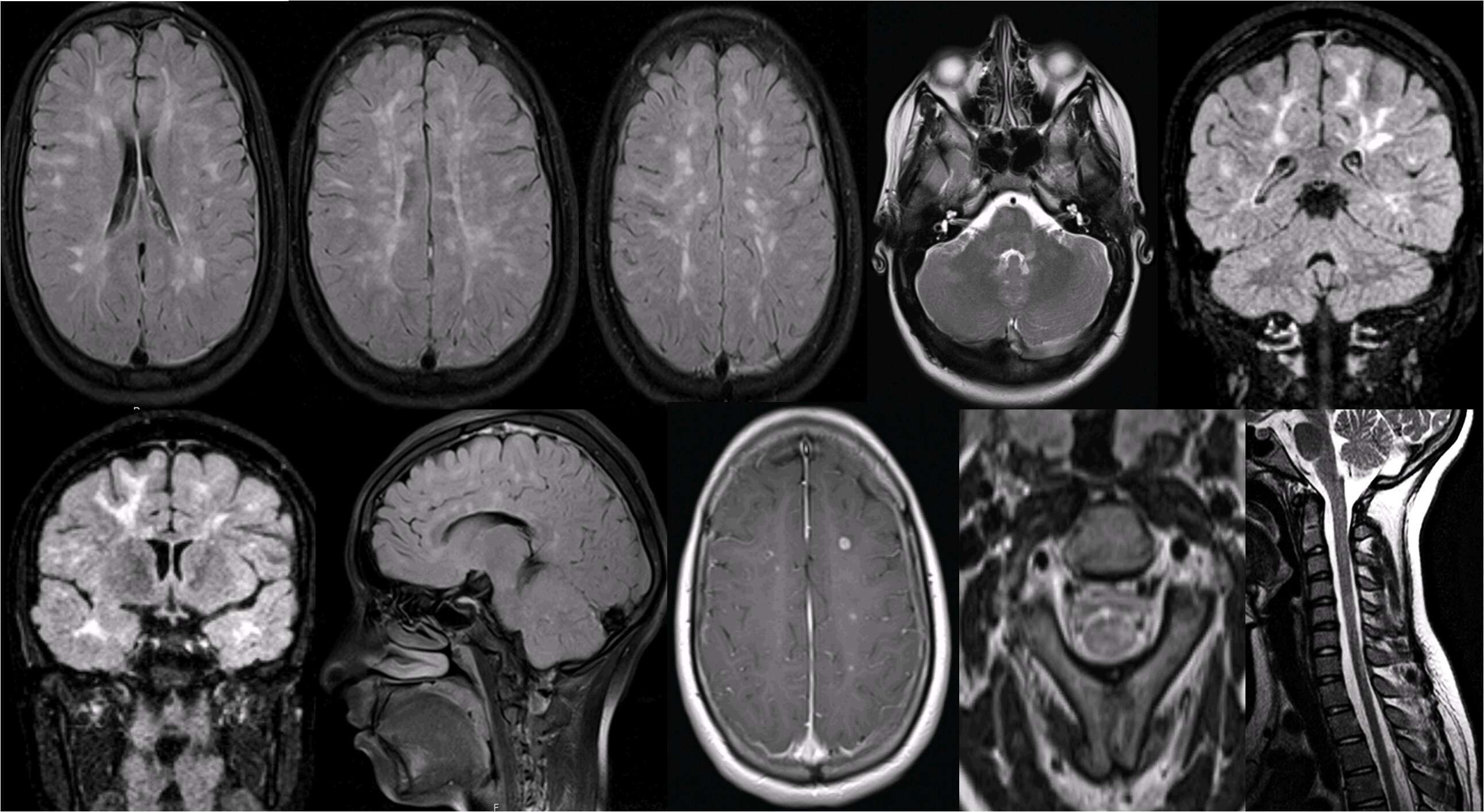
MRI showed countless multifocal injuries in the white matter, including juxtacortical, periventricular, subcortical, callosomarginal, capsular, in the protuberance, cerebellum and cervical spinal cord. To point out, coexisted injuries confluents of the white matter, subcortical and periventricular. Multiple lesions presented enhancement after administration of gadolinium.
Given the distribution in the temporal lobe, MS and CADASIL (cerebral autosomal dominant arteriopathy with subcortical infarcts and leukoencephalopathy) were considered as diagnostic hypotheses. However, the improvement after corticosteroids treatment and the presence of oligoclonal bands CSF helped confirm MS. The imaging findings meet the dissemination criteria in space and time (according to the criteria of McDonald Reviewed from 2017). Was initiated treatment with Natalizumab IV, with good results.
Conclusion: Despite the exuberant lesions on MRI, the wearer showed relatively little symptomatology and recovered completely after corticosteroids therapy. This case reminds us that imaging findings do not always reflect the clinical spectrum, highlighting the concept of clinical-radiological dissociation. It is also an atypical case since it is rare for pediatric MS to present itself with confluent white matter lesions in the initial phase of the disease.

Powered by Eventact EMS