
Teenage Girl with Chest Pain
Background: Pulmonary embolism is unusual in pediatric age. Clinical presentation may include chest pain and sudden onset dyspnea. It is often associated with the use of oral contraceptives, central venous catheters, trauma and hypercoagulable states. Its diagnosis requires a high degree of clinical suspicion.
Case Report: Previously healthy 16-year-old female, never a smoker, under oral contraception for 3 months, observed in the emergency department for sudden onset of pleuritic pain, unrelated to effort, for 48 hours. No history of trauma. No other respiratory symptoms, and no history of recent infections. Her mother had been diagnosed with portal vein thrombosis at the age of 32 (heterozygotic for the allelic variant MTHFR:C677T and homozygotic for PAI-1(4G)).
She was eupneic (SaO2 98-100%) and hemodynamically stable. Pulmonary and Cardiac auscultation were normal. No visible skins lesions.
Blood tests showed no elevation of inflammatory parameters, and D-Dimers 1381g/mL. PA Chest X-ray showed no signs of consolidation or pneumothorax.
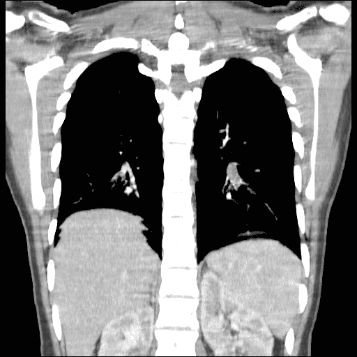
High-resolution thoracic CT scan with contrast revealed a filling defect seen in a branching subsegmental artery, that represented a clot. It was associated with a wedge shaped peripheral opacity of lung parenchyma in the left lower lobe as a result of lung infarction, secondary to pulmonary embolism.
She was admitted for anticoagulant therapy with enoxaparin (1mg/kg bid). Doppler echocardiogram and lower limb doppler ultrasound performed showed no relevant changes. She was discharged after 10 days, clinically well under oral anticoagulation. Currently awaits results for blood clotting disorders.
Conclusion: Particularly common in teenagers, chest pain is very often associated with anxiety, and represents a challenging symptom. With this case, the authors aim to present a rare case of pulmonary embolism in children, reinforcing the importance of a thorough clinical, family and personal history.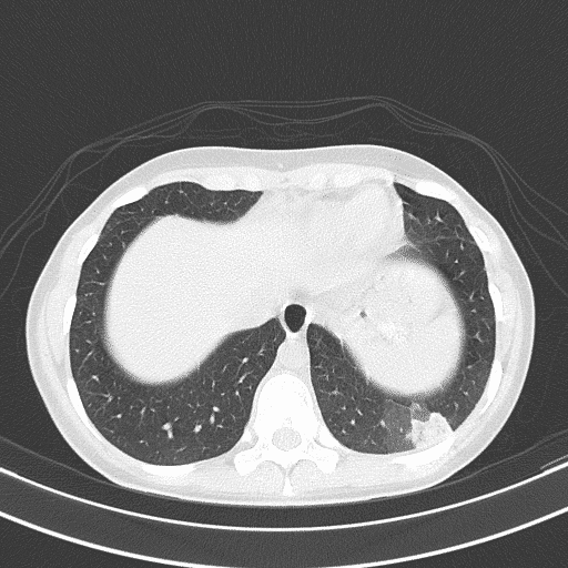
Powered by Eventact EMS