
Pediatric Cutaneous Horn of the Eyelid
2USF São João do Pragal, ACES Almada Seixal, Portugal
3Serviço de Pediatria, Hospital Garcia de Orta, Portugal
Background: A cutaneous horn consists of a keratotic projection that resembles a spicule or cone. Cutaneous horns of the eyelid are uncommon and even rarer in paediatric age. Typically, they grow in elder people with light skins and on sun-exposed areas. A variety of histopathological findings can be associated with this lesion.
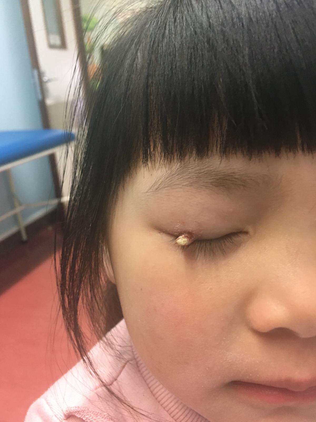
Clinical Case: A 4-year old girl presented to the urgency room with an upper eyelid lesion of the right eye. This lesion had 4 months of evolution. At examination she had no complains but pain with localized pressure. A minor haemorrhage happened the day before and motivated her consultation. This lesion had already been seen both at primary care center and general paediatric urgency before. Topical antibiotics and corticosteroids have been applied with no improvement. At this point, a referral was made to Ophthalmology. The clinical diagnosis of a cutaneous horn was made and she was sent to the ambulatory clinic for further investigation and excision biopsy.
Discussion: We present this case to highlight that, although most paediatric patients in Portugal are first seen by paediatricians or family doctors, there should be a low threshold for Ophthalmology referral in face of eyelid pathologies. Even though most of these lesions in children are benign, a careful management should be followed due to the possibility of a premalignant or malignant transformation at the lesion’s base.
Powered by Eventact EMS