")
LABEL-FREE REFRACTOMETRY AND PATHOGEN DETECTION BY BACK FOCAL PLANE IMAGING
2Biomedical Engineering, Technion – Israel Institute of Technology, Haifa, Israel
We present a new fluorescence microscopy method for label-free measurement of refractive index (RI) [1]. Our device is composed of a glass coverslip coated with a thin, dense layer of fluorophores inside a microfluidics channel. A high NA objective collects spatial frequencies which correspond to emission above the critical angle that is detectable if the fluorescent molecules are close to the coverslip – less than a wavelength away. The transition between the under-critical and super-critical fluorescence is linear in the conjugate back focal plane and corresponds to the change in medium RI according to Snell’s law. Imaging the back focal plane of the microscope directly allowed us to measure the RI with high precision by designing a custom algorithm and calibration scheme that are robust to various effects like photobleaching and tilt.
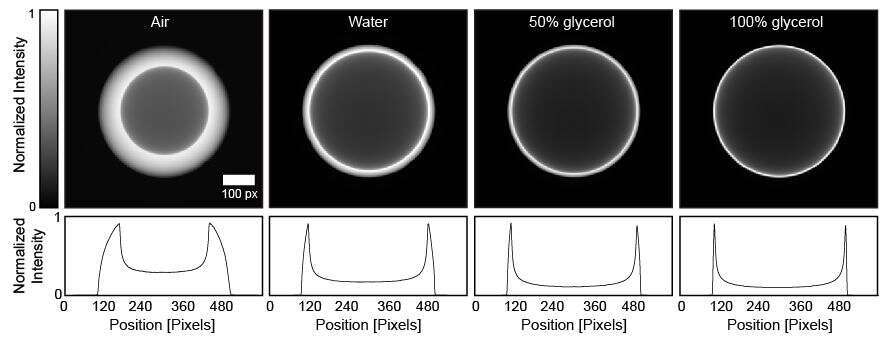
Figure 1. (Top) Experimentally measured back focal plane intensity images for two refractive indices and (Bottom) associated image cross-sections.
Our method enables label-free measurements of tiny amounts of liquid sample (picolitres of imaged volume) with high throughput and an order of magnitude higher precision than was previously reported over a large range of indices (1-1.45 in RI units). The sensitivity of our device enables detection of RI changes smaller than RI units, corresponding to a change of less than a 0.1% glycerol concentration in a water-glycerol solution. We furthermore demonstrate our system’s compatibility for biological applications, by measuring E. coli. growth in a chamber (bulk growth) for antibiotic resistance studies and single bacteria detection by scanning over a sparse sample with a narrow illumination beam.
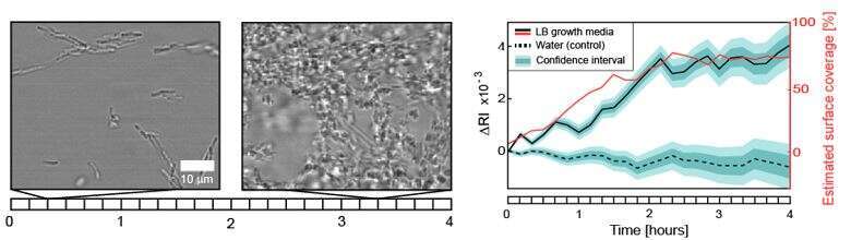
Figure 2. (Left) White light images of E. coli. growth at different times. (Right) The refractive index change corresponding to the growth over time.
- Ferdman, B., Weiss, L. E., Alalouf, O., Haimovich, Y., & Shechtman, Y. (2018). Ultrasensitive Refractometry via Supercritical Angle Fluorescence. ACS nano, 12(12), 11892-11898.
Powered by Eventact EMS