")
COMBINING LIGHT OPTICS WITH ELECTRON MICROSCOPY: LASER-STIMULATED ELECTRON ENERGY LOSS SPECTROSCOPY
2Material Science and Engineering, Technion – Israel Institute of Technology, Haifa, Israel
We will discuss new science and applications enabled by the ultrafast interactions of electrons and laser pulses inside electron microscopes. Such interactions enable novel microscopy techniques with time-correlated measurements (Fig.1). In particular, we demonstrate a new method of laser-stimulated electron energy loss spectroscopy (LS-EELS). As a result of the stimulated laser excitation, LS-EELS can increase the contrast of conventional EELS by order of magnitude and improve its energy resolution. Therefore, LS-EELS can give more complete information on the electric and optical properties of materials, beyond the capabilities of conventional EELS. We will present our recent study of a photonic crystal (PhC) structure (Fig.2a) where we measured the complete band structure map (Fig.2c) and imaged the different modes (Fig.2b) with subwavelength resolution. We show how the PhC structures enables strong interactions with relatively low laser power, thus it can serve as a platform for future LS-EELS experiments, for example to enable probing the excited states of a single molecule with improved energy resolution and contrast. Ultimately, this method could be improved towards the molecular scale to enable optical spectroscopy with sub-meV energy resolution and sub-nm spatial resolution.
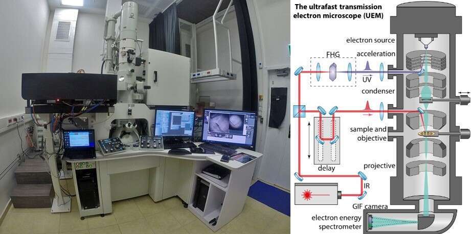
Figure 1: The ultrafast transmission electron microscope (UTEM) at the Technion. Left: Image of the newly-installed setup. Right: Illustration of the UTEM operation: showing the interaction of ultrafast electron pulses (green) and laser pulses (red) with a TEM grid. The results of such interactions can be inferred by electron energy loss spectroscopy (EELS) with energy-filtered imaging (bottom of the column).
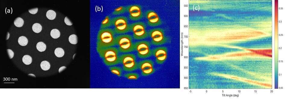
Figure 2: Preliminary results of Imaging light with a TEM, demonstrating the Laser-stimulated EELS technique. (a) A photonic crystal (PhC) slab: 200nm thick SiN membrane with periodic holes . (b) Energy-filtered image of the PhC during laser illumination. Laser-stimulated EELS enables imaging of the resonant coupling of the laser into a specific resonant (Bloch) mode of light trapped in the membrane. (c) Mapping the PhC bandstructure, with each pixel found through a separate laser-stimulated EELS experiment. Red colors represent efficient coupling to particular PhC resonant (Bloch) modes.
Powered by Eventact EMS