
Plasticity in Au crystals studied by In-situ nano-indentation coupled with bragg coherent diffraction imaging
2Materials Science and Engineering, Technion – Israel Institute of Technology, Haifa
The mechanical properties of micro- and nanostructures were demonstrated to vary significantly from their bulk counterparts. Despite numerous studies, plasticity at the nanoscale is, however, not fully understood yet. In-situ experiments are perfectly suited for the fundamental understanding of the onset of dislocation nucleation. Here, we combined in-situ nano-indentation of single-crystalline as well as twinned Au particles with Bragg Coherent Diffraction Imaging (BCDI) that is non-invasive X-ray technique and that is highly sensitive to both strain and defects. Previous BCDI studies on indented Au crystals demonstrated the capability to imaging a single prismatic loop induced by nano-indentation and trapped inside the crystal [1]. Since any movement of diffractometer motors may induce vibrations that eventually lead to damaging the nano-crystal under load, ordinary rocking scans are not suitable for recording 3D reciprocal space maps in-situ. Thanks to the achromacity of the KB mirrors, we scanned the energy of the incident X-ray beam, thus probing the intensity distribution in reciprocal space at different loading steps, thus imaging the evolution of strain and defects (see Fig. 1).
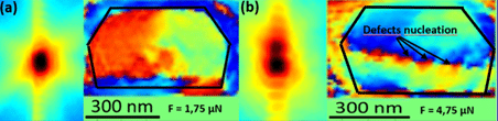
Figure 1: a,b) Qz-Qy integrated images of the core of 3D diffraction patterns for two different mechanical loads and Z-Y cut of reconstructions of the phase for the corresponding mechanical loads.
To the best of our knowledge, this is the first time that E-BCDI has been successfully employed during in-situ experiments providing direct insight into the plasticity at the nanoscale and, in particular, the onset of defect nucleation.
References
[1] - M. Dupraz, G. Beutier, TW. Cornelius, G. Parry, Z. Ren, S. Labat, M.-I. Richard, G.A. Chahine, O. Kovalenko, M. De Boissieu, E. Rabkin, M. Verdier, O. Thomas, Nano Lett. 17, 6696 (2017)

Powered by Eventact EMS