
Three-dimensional structural imaging of defects in Pt nanocrystals
2Inorganic Materials & Catalysis, Eindhoven University of Technology, Eindhoven
3Materials Science and Engineering, Technion Israel Institute of Technology, Haifa
4Experimental division, The European Synchrotron, Grenoble
At the nanoscale, the properties of materials are largely influenced by elastic strain and depend critically on the presence of crystal defects. However, imaging and characterising the structure of defects inside a crystal in three-dimensions (3D) and in situ during reaction remain a challenge. Here, we demonstrate the capabilities of Bragg coherent diffraction imaging [1] to reveal in 3D the structure of defects in Platinum (Pt) nanocrystals and their associated lattice strains. Dislocations are characterised from their characteristic displacement and strain fields (see Figure 1, [2]). We also succeeded to reveal in 3D the detwinning process in a single Pt nanoparticle during in situ gas reaction while increasing the O2 partial pressure [3]. In situ and non-invasive structural characterisation of defects during reaction opens new avenues for understanding defect behaviors in confined crystals and paves the way for strain and defect engineering.
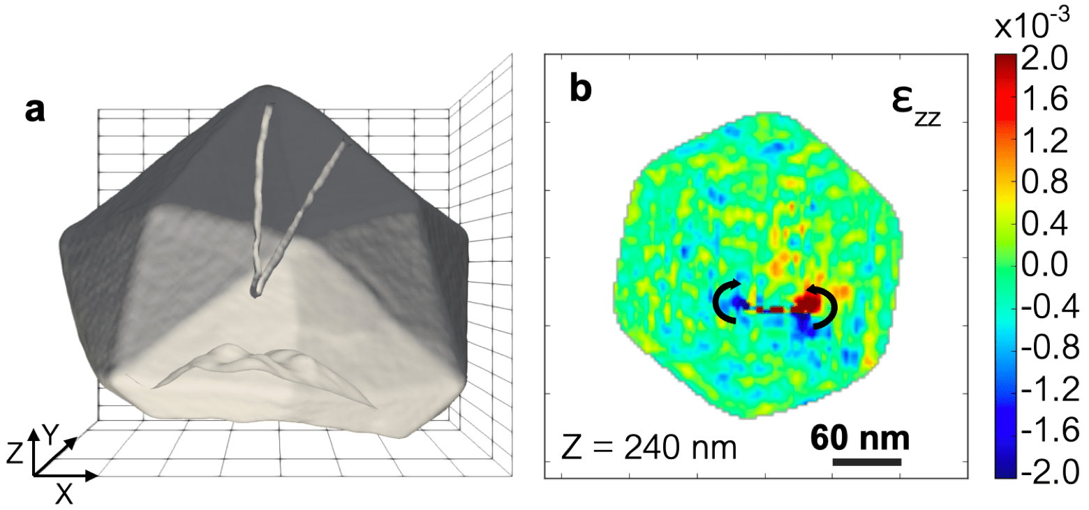
Figure 1: (a) Wireframe plot of the reconstructed electron density of a Pt particle (diameter of 350 nm) drawn at 35% of the maximum density. A dislocation loop is evidenced. (b) Two-dimensional cut of the out-of-plane strain, ezz, at a particle height of 240 nm.
[1] S. Labat, M.-I. Richard, M. Dupraz, M. Gailhanou, G. Beutier, M. Verdier, F. Mastropietro, T. W. Cornelius, T. U. Schülli, J. Eymery, and O. Thomas, ACS Nano 9, 9210 (2015).
[2]-[3] To be submitted.

Powered by Eventact EMS