")
STAR OF DAVID
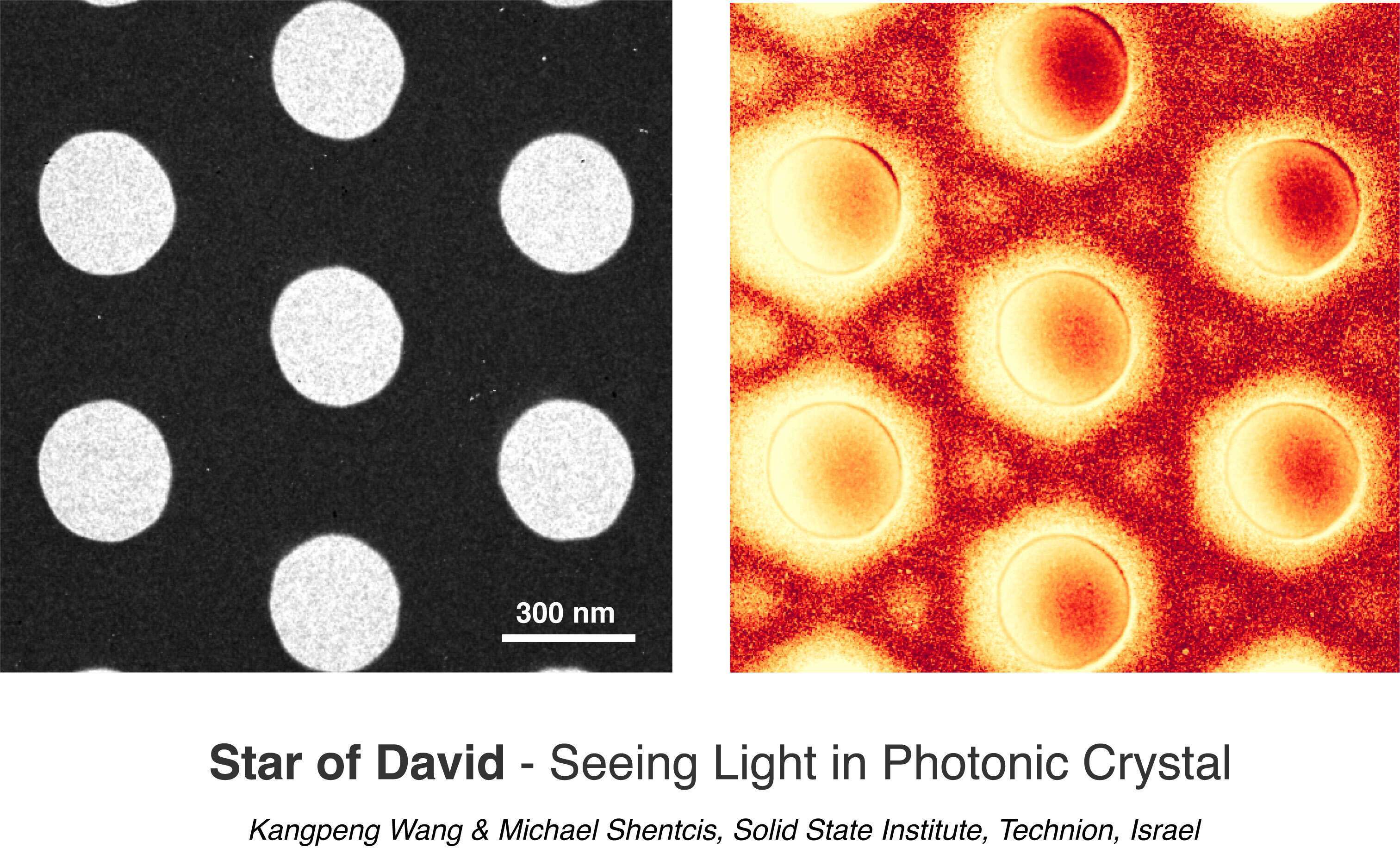
The first direct subwavelength imaging of light trapped in a photonic crystal (PhC) slab. Electrons are used to image the electric field of a laser light pulse by synchronizing their arrival time with the laser pulse in an ultrafast transmission electron microscope (UTEM). The PhC is a 200 nm thick Si3N4 membrane with 300 nm holes and 600 nm periodicity in triangular geometry (left mono-color TEM image). The UTEM enables novel microscopy techniques with femtosecond temporal and nanometer spatial resolutions. Through the quantized electron-laser interaction, each electron can absorb or emit an integer number of photons from the driving laser pulse, such that the electron energy spectrum contains discrete peaks on both sides of the zero-loss peak. Consequently, we image the laser light in the sample via energy-filtered transmission electron microscopy (EFTEM), filtering on the regime of energy gain (right pseudo-color image). The PhC is excited by a 624 nm, 250 fs laser, incident at ~5 degrees in the Gamma-M direction to the PhC slab.
Powered by Eventact EMS