")
GRUMPY FROG
Molecular Cell Biology, Weizmann Institute of Science, Rehovot, Israel
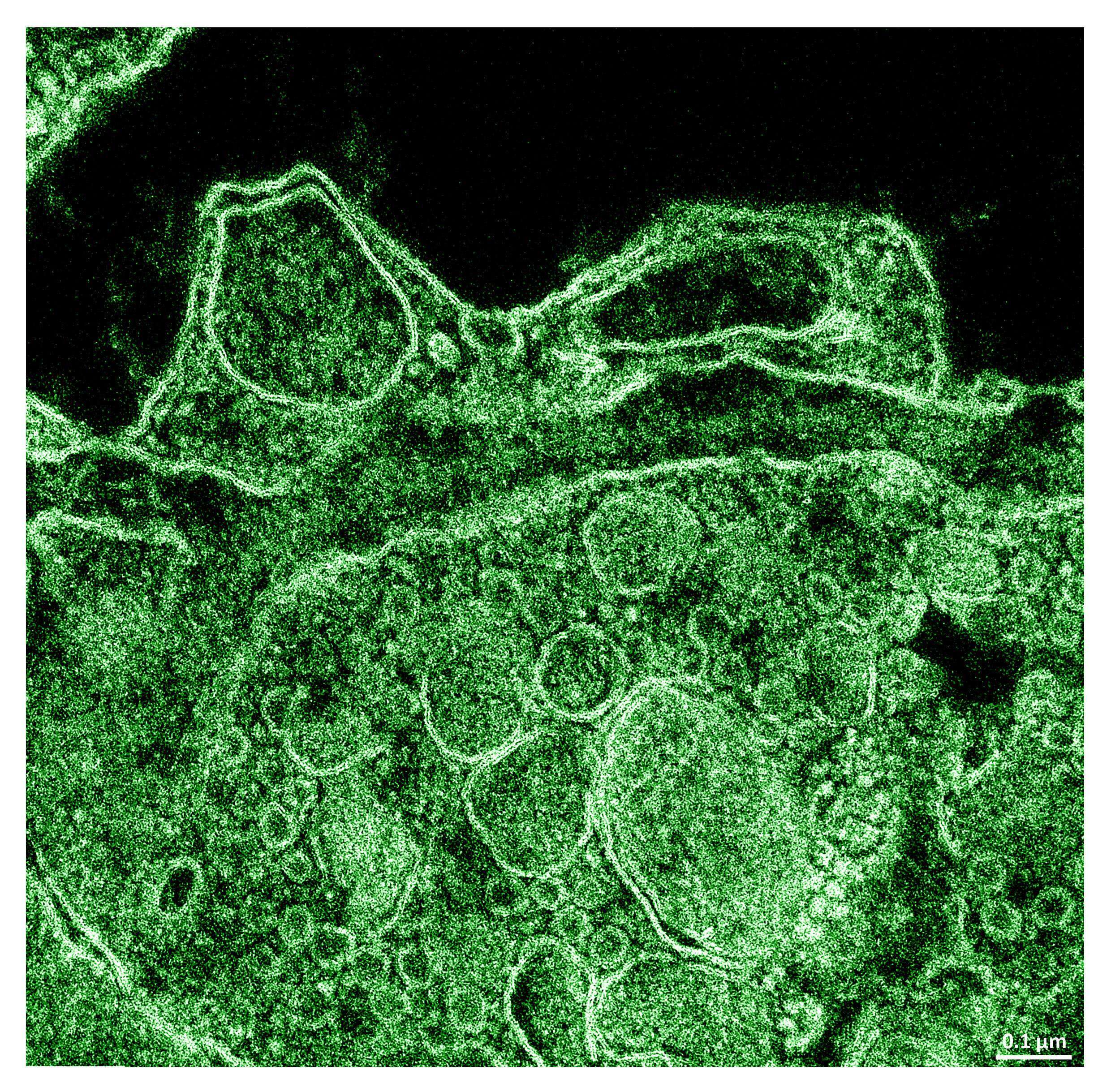
This image shows the contact between the hormone-secretory cell and blood capillary separated by perivascular space. The ‘frog’ is depicted by two adjacent macropinocytic vesicles of the endothelial cell – the eyes; and by perivascular space – the mouth. The image was taken with transmission electron microscope, following chemical fixation of the whole pituitary gland dissected from adult fish.
Powered by Eventact EMS